


1. Introduction and Methodology
Sponsored by: Congress of Neurological Surgeons and the Section on Disorders of the Spine and Peripheral Nerves in collaboration with the Section on Neurotrauma and Critical Care
Endorsed by: The Congress of Neurological Surgeons (CNS) and the American Association of Neurological Surgeons (AANS)
John E. O’Toole, MD, MS1, Michael G. Kaiser, MD,2, Paul A. Anderson, MD3, Paul M. Arnold, MD4, John H. Chi, MD, MPH5, Andrew T. Dailey, MD6, Sanjay S. Dhall, MD7, Kurt M. Eichholz, MD8 , James S. Harrop, MD9, Daniel J. Hoh, MD10, Sheeraz Qureshi, MD, MBA11, Craig H. Rabb, MD12, and P. B. Raksin, MD13
1Department of Neurological Surgery, Rush University Medical Center, Chicago, Illinois
2Department of Neurosurgery, Columbia University, New York, New York
3Department of Orthopedics and Rehabilitation, University of Wisconsin, Madison, Wisconsin
4Department of Neurosurgery, University of Kansas School of Medicine, Kansas City, Kansas
5Department of Neurosurgery, Harvard Medical School, Brigham and Women’s Hospital, Boston, Massachusetts
6Department of Neurosurgery, University of Utah, Salt Lake City, Utah
7Department of Neurological Surgery, University of California, San Francisco, San Francisco, California
8St. Louis Minimally Invasive Spine Center, St. Louis, Missouri
9Departments of Neurological Surgery and Orthopedic Surgery, Thomas Jefferson University, Philadelphia, Pennsylvania
10Lillian S. Wells Department of Neurological Surgery, University of Florida, Gainesville, Florida
11Department of Orthopaedic Surgery, Weill Cornell Medical College, New York, New York
12Department of Neurosurgery, University of Utah, Salt Lake City, Utah
13Division of Neurosurgery, John H. Stroger, Jr. Hospital of Cook County and Department of Neurological Surgery, Rush University Medical Center, Chicago, Illinois
Correspondence:
John E. O’Toole, MD, MS
Professor of Neurosurgery
Rush University Medical Center
1725 West Harrison St. Suite 855
Chicago, IL 60612
E-mail: john_otoole@rush.edu
Keywords: clinical practice guideline, lumbar fracture, thoracic fracture, thoracolumbar fracture
No part of this manuscript has been published or submitted for publication elsewhere.
Disclaimer of Liability
This clinical systematic review and evidence-based guideline was developed by a multidisciplinary physician volunteer task force and serves as an educational tool designed to provide an accurate review of the subject matter covered. These guidelines are disseminated with the understanding that the recommendations by the authors and consultants who have collaborated in their development are not meant to replace the individualized care and treatment advice from a patient’s physician(s). If medical advice or assistance is required, the services of a competent physician should be sought. The proposals contained in these guidelines may not be suitable for use in all circumstances. The choice to implement any particular recommendation contained in these guidelines must be made by a managing physician in light of the situation in each particular patient and on the basis of existing resources.
ABSTRACT
Background: The thoracic and lumbar (“thoracolumbar”) spine are the most commonly injured region of the spine in blunt trauma. Trauma of the thoracolumbar spine is frequently associated with spinal cord injury and other visceral and bony injuries. Prolonged pain and disability after thoracolumbar trauma present a significant burden on patients and society.
Objective: To formulate evidence-based clinical practice recommendations for the care of patients with injuries to the thoracolumbar spine.
Methods: A systematic review of the literature was performed using the National Library of Medicine PubMed database and the Cochrane Library for studies relevant to thoracolumbar spinal injuries based on specific clinically oriented questions. Relevant publications were selected for review.
Results: For all of the questions posed, the literature search yielded a total of 6561 abstracts. The task force selected 804 articles for full text review, and 78 were selected for inclusion in this overall systematic review.
Conclusion: The available evidence for the evaluation and treatment of patients with thoracolumbar spine injuries demonstrates considerable heterogeneity and highly variable degrees of quality. However, the workgroup was able to formulate a number of key recommendations to guide clinical practice. Further research is needed to counter the relative paucity of evidence that specifically pertains to patients with only thoracolumbar spine injuries.
INTRODUCTION
Goals and Rationale
Traumatic injuries of the thoracic and lumbar spine (“thoracolumbar”) occur in approximately 7% of all blunt trauma patients and comprise 50% to 90% of the 160,000 annual traumatic spinal fractures in North America.1–5 Up to 25% of patients with thoracolumbar fractures have concomitant spinal cord injury.1,4 Long-term care of patients with persistent disability after thoracolumbar trauma represents a significant burden on society’s health care resources.1,2,4–6 In addition, these patients frequently have multiple visceral and bony injuries, and treatment decision-making can be quite challenging4,6 For the purposes of this guideline, “thoracolumbar” includes the distinct regions of the rigid thoracic spine (T1-10), transitional thoracolumbar junction (T10-L2), and flexible lumbar spine (L3-5). Patients with injuries to these various spinal regions are frequently studied together; therefore, this pooling is needed to more successfully assess the content of the evidence base and allow for practical clinical implementation of the recommendations formulated. The rationale for examining injuries of the thoracolumbar spine separate from injuries of cervical spine lies in the distinct anatomic, physiologic, and epidemiologic characteristics of these 2 groups of patients. Thoracolumbar injuries are more common, concomitant visceral injuries are more frequently seen in thoracolumbar trauma, the types of fractures encountered are anatomically unique, the vascular supply of the thoracic spinal cord is more tenuous than the cervical spinal cord, and surgical decision-making and surgical procedures are different in the two populations.1–6
There remains a lack of consensus on a number of issues surrounding the care of these patients, including 1) formal injury classification systems; 2) appropriate radiological evaluation; 3) neurological assessment instrument; 4) systemic treatments for blood pressure and spinal cord injury management; 5) venous thromboembolism prophylaxis; 6) nonoperative versus operative management; 7) choice of surgical approach; 8) timing of surgical treatment; and 9) novel surgical techniques.1–3,5–11The American Association of Neurological Surgeons (AANS)/Congress of Neurological Surgeons (CNS) Section on Disorders of the Spine and Peripheral Nerves and the Section on Neurotrauma and Critical Care initiated this effort to develop a clinical practice guideline to answer key clinical questions regarding the care of patients with thoracolumbar trauma using the available evidence base and employing a rigorous guideline elaboration methodology. In particular, specific patient, intervention, comparison, outcome (PICO) questions were posed that the workgroup and sponsoring sections felt were the most pertinent to, and in some cases the most controversial, in the routine clinical care of these patients. These PICO questions were formulated before commencing with any literature search or evidence abstraction.
Ultimately, this clinical guideline was created to improve patient care by outlining the appropriate information gathering and decision-making processes involved in the evaluation and treatment of patients with thoracolumbar spine trauma. The surgical management of these patients often takes place under a variety of circumstances and by various clinicians. This guideline was created as an educational tool to guide qualified physicians through a series of diagnostic and treatment decisions in an effort to improve the quality and efficiency of care.
METHODS
The guidelines task force initiated a systematic review of the literature relevant to the diagnosis and treatment of patients with thoracolumbar trauma. Through objective evaluation of the evidence and transparency in the process of making recommendations, this evidence-based clinical practice guideline was developed for the diagnosis and treatment of adult patients with thoracolumbar injury. These guidelines are developed for educational purposes to assist practitioners in their clinical decision-making processes.
Literature Search
The task force members identified search terms/parameters, and a medical librarian implemented the literature search, consistent with literature search protocols, using the National Library of Medicine PubMed database and the Cochrane Library (which included the Cochrane Database of Systematic Reviews, the Database of Abstracts of Reviews of Effect, the Cochrane Central Register of Controlled Trials, the Health Technology Assessment Database, and the National Health Service Economic Evaluation Database) for the period from January 1, 1946 to March 31, 2015, using the search strategies provided in each paper of this guideline series.
RESULTS
A total of 6561 studies were yielded from the literature searches for all chapters of this guideline. Task force members reviewed all abstracts yielded from the literature search and identified a total of 804 studies for full text review and extraction, addressing the clinical questions in accordance with the literature search protocols (provided in each chapter). Task force members identified the best research evidence available to answer the targeted clinical questions. When level I, II, or III literature was available to answer specific questions, the task force did not review level IV studies. A total of 78 studies were selected for inclusion in the systematic review (Appendix I). Selection of studies for full-text review were conducting by 2 members of the workgroup (unique for each chapter) in conjunction with CNS Guidelines staff. Disagreements regarding selection of abstracts for full-text review were resolved by consensus under the guidance of the workgroup chairs.
Inclusion/Exclusion Criteria
Articles were retrieved and included only if they met specific inclusion/exclusion criteria. These criteria were also applied to articles provided by guideline task force members who supplemented the electronic database searches with articles from their own files. To reduce bias, these criteria were specified before conducting the literature searches.
Articles that did not meet the following criteria, for the purposes of this evidence-based clinical practice guideline, were excluded. To be included as evidence in the guideline, an article had to be a report of a study that:
- Investigated patients with thoracolumbar injuries;
- Included patients ≥18 years of age;
- Enrolled ≥80% of thoracolumbar injuries (studies with mixed patient populations were included if they reported results separately for each group/patient population);
- Was a full article report of a clinical study;
- Was not an internal medical records review, meeting abstract, historical article, editorial, letter, or commentary;
- Appeared in a peer-reviewed publication or a registry report;
- Enrolled ≥10 patients per arm per intervention (20 total) for each outcome;
- Included only human subjects;
- Was published in or after 1946;
- Quantitatively presented results;
- Was not an in vitro study;
- Was not a biomechanical study;
- Was not performed on cadavers;
- Was published in English;
- Was not a systematic review, meta-analysis, or guideline developed by others*
- Was a case series (therapeutic study) where higher level evidence exists.
Rating Quality of Diagnostic Evidence
The guideline task force used a modified version of the North American Spine Society’s evidence-based guideline development methodology.12The North American Spine Society methodology uses standardized levels of evidence (Appendix II) and grades of recommendation (Appendix III) to assist practitioners in easily understanding the strength of the evidence and the recommendations within the guidelines. The levels of evidence range from level I (high-quality randomized controlled trial) to level IV (case series). Grades of recommendation indicate the strength of the recommendations made in the guideline based on the quality of the literature. Levels of evidence have specific criteria and are assigned to studies before developing recommendations. Recommendations are then graded based upon the level of evidence. To better understand how levels of evidence inform the grades of recommendation and the standard nomenclature used within the recommendations, see Appendix III.
Guideline recommendations were written using a standard language that indicates the strength of the recommendation. “A” recommendations indicate a test or intervention is “recommended”; “B” recommendations “suggest” a test or intervention; “C” recommendations indicate that a test or intervention or “is an option.” “Insufficient evidence” statements clearly indicate that “there is insufficient evidence to make a recommendation for or against” a test or intervention. Task force consensus statements clearly state that “in the absence of reliable evidence, it is the task force’s opinion that” a test or intervention may be considered. Both the levels of evidence assigned to each study and the grades of each recommendation were arrived at by consensus of the workgroup using up to three rounds of voting when necessary.
In evaluating studies as to levels of evidence for this guideline, the study design was interpreted as establishing only a potential level of evidence. For example, a therapeutic study designed as a randomized controlled trial would be considered a potential level I study. The study would then be further analyzed as to how well the study design was implemented, and significant shortcomings in the execution of the study would be used to downgrade the levels of evidence for the study’s conclusions (see Appendix IV for additional information and criteria).
Revision Plans
In accordance with the Institute of Medicine’s standards for developing clinical practice guidelines and criteria specified by the National Guideline Clearinghouse, the task force will monitor related publications after the release of this document and will revise the entire document and/or specific sections “if new evidence shows that a recommended intervention causes previously unknown substantial harm; that a new intervention is significantly superior to a previously recommended intervention from an efficacy or harms perspective; or that a recommendation can be applied to new populations.”13 In addition, the task force will confirm within 5 years from the date of publication that the content reflects current clinical practice and the available technologies for the evaluation and treatment for patients with thoracolumbar trauma.
DISCUSSION
This guideline yields a number of important recommendations but also highlights the paucity of high-level evidence regarding the care of specifically the thoracolumbar trauma population. Many (if not most) spinal trauma publications contain a high degree of heterogeneity in the populations studied, including levels of injury (mixed cervical and thoracolumbar), mechanisms of injury, anatomical classifications of injury, neurological status at admission, interventions performed, outcomes studied, and follow-up periods. For this guideline, the evidence base allowed only one grade A recommendation: “Surgeons should understand that the addition of arthrodesis to instrumented stabilization has not been shown to impact clinical or radiologic outcomes, and adds to increased blood loss and operative time.” Several grade B and C recommendations were also made, but in many circumstances, the workgroup was left with inconclusive evidence to answer the salient clinical questions posed. In two cases, the workgroup felt compelled to generate recommendations based solely on consensus because of what was perceived as current standard of care with minimal risk posed to the patient (blood pressure management and venous thromboembolism prophylaxis).
Future Research
Each chapter within this guideline explores areas of need for future research for each topic of interest. However, as a general need, future studies should attempt to analyze patients with thoracolumbar injuries separate from those with cervical injuries to better clarify the most effective diagnostic and treatment modalities for these patients in particular.
CONCLUSION
Ultimately, this clinical practice guideline serves as a critical reference for clinicians caring for adult patients with thoracolumbar trauma. This synthesis of the most contemporary evidence using rigorous methodology provides the reader with an important resource to address key questions in routine clinical practice. As with all evidence-based guidelines, however, it should be implemented in conjunction with clinician expertise and patient preferences.
Potential Conflicts of Interest
The task force members were required to report all possible conflicts of interest (COIs) before beginning work on the guideline, using the COI disclosure form of the AANS/CNS Joint Guidelines Review Committee, including potential COIs that are unrelated to the topic of the guideline. The CNS Guidelines Committee and Guideline Task Force Chairs reviewed the disclosures and either approved or disapproved the nomination. The CNS Guidelines Committee and Guideline Task Force Chairs are given latitude to approve nominations of Task Force members with possible conflicts and address this by restricting the writing and reviewing privileges of that person to topics unrelated to the possible COIs. The conflict of interest findings are provided in detail in Appendix V.
ACKNOWLEDGEMENTS
The guidelines task force would like to acknowledge the CNS Guidelines Committee for their contributions throughout the development of the guideline and the AANS/CNS Joint Guidelines Review Committee for their review, comments, and suggestions throughout peer review, as well as the contributions of Trish Rehring, MPH, CHES, Senior Manager of Clinical Practice Guidelines for the CNS, and Mary Bodach, MLIS, Guidelines Specialist and Medical Librarian for assistance with the literature searches. Throughout the review process the reviewers and authors were blinded from one another. At this time, the guidelines task force would like to acknowledge the following individual peer reviewers for their contributions: Maya Babu, MD, MBA, Greg Hawryluk, MD, PhD, Steven Kalkanis, MD, Yi Lu, MD, PhD, Jeffrey J. Olson, MD, Martina Stippler, MD, Cheerag Upadhyaya, MD, MSc, and Robert Whitmore, MD.
Disclosures
These evidence-based clinical practice guidelines were funded exclusively by the Congress of Neurological Surgeons and the Section on Disorders of the Spine and Peripheral Nerves in collaboration with the Section on Neurotrauma and Critical Care, which received no funding from outside commercial sources to support the development of this document.
Appendix I. Article Inclusions and Exclusions
Included and Excluded Articles Flowchart

Appendix II. Rating Evidence Quality
Levels of Evidence for Primary Research Questiona
Therapeutic studies – Investigating the results of treatment
| Types of studies | ||||
|---|---|---|---|---|
| Therapeutic studies – Investigating the results of treatment | Prognostic studies – Investigating the effect of a patient characteristic on | Diagnostic studies – Investigating a diagnostic test | Economic and decision analyses – Developing an economic or decision | |
| Level I | ||||
| High-quality randomized trial with statistically significant difference or no statistically significant difference but narrow confidenceintervals Systematic reviewb of level I RCTs (and study results were homogenousc) | High-quality prospective studyd (all patients were enrolled at the same point in their disease with ≥80% follow-up of enrolled patients) Systematic reviewb of level I studies | Testing of previously developed diagnostic criteria on consecutive patients (with universally applied reference “gold” standard) Systematic reviewb of level I studies | Sensible costs and alternatives; values obtained from many studies; with multiway sensitivity analyses Systematic reviewb of level I studies | |
| Level II | ||||
| Lesser quality RCT (e.g., ≤80% follow-up, no blinding, or improper randomization) Prospectived comparative studye Systematic reviewb of level II studies or level I studies with inconsistent results | Retrospectivef study Untreated controls from an RCT Lesser quality prospective study (e.g., patients enrolled at different points in their disease or ≤80% follow-up) Systematic reviewb of level II studies | Development of diagnostic criteria on consecutive patients (with universally applied reference “gold” standard) Systematic reviewb of level II studies | Sensible costs and alternatives; values obtained from limited studies; with multiway sensitivity analyses Systematic reviewb of level II studies | |
| Level III | ||||
| Case control studyg Retrospectivef comparative studye Systematic reviewb of level III studies | Case control studyg | Study of nonconsecutive patients; without consistently applied reference “gold” standard Systematic reviewb | Analyses based on limited alternatives and costs; and poor estimates Systematic reviewb of level III studies | |
| Level IV | ||||
| Case seriesh | Case series | Case-control study Poor reference standard | Analyses with no sensitivity analyses |
RCT, Randomized controlled trial.
aA complete assessment of quality of individual studies requires critical appraisal of all aspects of the study design.
bA combination of results from ≥2 previous studies.
cStudies provided consistent results.
dStudy was started before the first patient enrolled.
ePatients treated one way (e.g., instrumented arthrodesis) compared with a group of patients treated in another way (e.g., unsintrumented arthrodesis) at the same institution.
fThe study was started after the first patient enrolled. gPatients identified for the study based on their outcome, called “cases” (e.g., pseudoarthrosis) are compared to those who did not have outcome, called “controls” (e.g., successful fusion).
hPatients treated one way with no comparison group of patients treated in another way.
Appendix III. Linking Levels of Evidence to Grades of Recommendation
| Grade of recommendation | Standard language | Levels of evidence | |
|---|---|---|---|
| A | Recommended | Two or more consistent level I studies | |
| B | Suggested | One level I study with additional supporting level II or III studies | Two or more consistent level II or III studies |
| C | Is an option | One level I, II, or III study with supporting level IV studies | Two or more consistent level IV studies |
| Insufficient (insufficient or conflicting evidence) | Insufficient evidence to make recommendation for or against | A single level I, II, III, or IV study without other supporting evidence | >1 study with inconsistent findingsa |
aNote that in the presence of multiple consistent studies, and a single outlying, inconsistent study, the Grade of Recommendation will be based on the level of the consistent studies.
Appendix IV. Criteria Grading the Evidence
The task force used the criteria provided below to identify the strengths and weaknesses of the studies included in this guideline. Studies containing deficiencies were downgraded one level (no further downgrading allowed, unless so severe that study had to be excluded). Studies with no deficiencies based on study design and contained clinical information that dramatically altered current medical perceptions of topic were upgraded.
1. Baseline study design (i.e., therapeutic, diagnostic, prognostic) determined to assign initial level of evidence.
2. Therapeutic studies reviewed for following deficiencies:
- Failure to provide a power calculation for an RCT;
- High degree of variance or heterogeneity in patient populations with respect to presenting diagnosis/demographics or treatments applied;
- <80% of patient follow-up;
- Failure to utilize validated outcomes instrument;
- No statistical analysis of results;
- Cross over rate between treatment groups of >20%;
- Inadequate reporting of baseline demographic data;
- Small patient cohorts (relative to observed effects);
- Failure to describe method of randomization;
- Failure to provide flowchart following patients through course of study (RCT);
- Failure to account for patients lost to follow-up;
- Lack of independent post-treatment assessment (e.g., clinical, fusion status, etc.);
- Utilization of inferior control group:
- Historical controls;
- Simultaneous application of intervention and control within same patient.
- Failure to standardize surgical/intervention technique;
- Inadequate radiographic technique to determine fusion status (e.g., static radiographs for instrumented fusion).
3. Methodology of diagnostic studies reviewed for following deficiencies:
- Failure to determine specificity and sensitivity;
- Failure to determine inter- and intraobserver reliability;
- Failure to provide correlation coefficient in the form of kappa values.
4. Methodology of prognostic studies reviewed for following deficiencies:
- High degree of variance or heterogeneity in patient populations with respect to presenting diagnosis/demographics or treatments applied;
- Failure to appropriately define and assess independent and dependent variables (e.g., failure to use validated outcome measures when available).
Appendix V. Conflicts of Interest>
| Author | Potential COI |
|---|---|
| Paul A. Anderson, MD | Aesculap: Consultant fee SI Bone: Stock shareholder Spartec: Stock shareholder Expanding Orthopedics: Stock shareholder Titan Spine: Stock shareholder RTI: Other financial support Stryker: Other financial support Lumbar Spine Research Society: Board, trustee or officer position (President) |
| Paul M. Arnold, MD | Medtronic: Consultant fee Sofamor Danek: Consultant fee Spine Wave: Consultant fee InVivo: Consultant fee Stryker Spine: Consultant fee Evoke Medical: Stock shareholder Z-Plasty: Stock shareholder AO Spine North America: Sponsored or reimbursed travel (for self only) |
| John H. Chi, MD, MPH | DePuy Spine: Consultant fee K2M: Consultant fee |
| Andrew Dailey, MD | K2M: Grants/Research support K2M: Consultant fee Zimmer Biomet: Consultant fee Medtronic: Consultant fee |
| Sanjay Dhall, MD | Globus Medical: Honorarium Depuy Spine: Honorarium |
| James S. Harrop, MD | DePuy Spine: Consultant fee Asterias: Other financial support/Scientific advisor Tejin: Other financial support/Scientific advisor Bioventus: Other financial support/Scientific advisor AO Spine: Board, trustee, or officer position |
| John E. O’Toole, MD, MS | Globus Medical: Consultant fee RTI Surgical: Consultant fee Theracell, Inc.: Stock shareholder |
REFERENCES
1. Ghobrial GM, Jallo J. Thoracolumbar spine trauma: Review of the evidence. J Neurosurg Sci 2013;57:115-122.
2. Ghobrial GM, Maulucci CM, Maltenfort M, et al. Operative and nonoperative adverse events in the management of traumatic fractures of the thoracolumbar spine: a systematic review. Neurosurg Focus 2014;37:E8.
3. Joaquim AF, Patel AA. Thoracolumbar spine trauma: Evaluation and surgical decision-making. J Craniovertebr Junction Spine 2013;4:3-9.
4. Katsuura Y, Osborn JM, Cason GW. The epidemiology of thoracolumbar trauma: A meta-analysis. J Orthop 2016;13:383-388.
5. Wood KB, Li W, Lebl DR, Ploumis A. Management of thoracolumbar spine fractures. Spine J 2014;14:145-164.
6. Dai LY, Yao WF, Cui YM, Zhou Q. Thoracolumbar fractures in patients with multiple injuries: Diagnosis and treatment-a review of 147 cases. J Trauma 2004;56:348-355.
7. Fu MC, Nemani VM, Albert TJ. Operative treatment of thoracolumbar burst fractures: Is fusion necessary? HSS J 2015;11:187-189.
8. Gertzbein SD. Scoliosis Research Society. Multicenter spine fracture study. Spine (Phila Pa 1976) 1992;17:528-540.
9. Lopez AJ, Scheer JK, Smith ZA, Dahdaleh NS. Management of flexion distraction injuries to the thoracolumbar spine. J Clin Neurosci 2015;22:1853-1856.
10. Scheer JK, Bakhsheshian J, Fakurnejad S, Oh T, Dahdaleh NS, Smith ZA. Evidence-based medicine of traumatic thoracolumbar burst fractures: A systematic review of operative management across 20 years. Global Spine J 2015;5:73-82.
11. Schroeder GD, Harrop JS, Vaccaro AR. Thoracolumbar trauma classification. Neurosurg Clin N Am 2017;28:23-29.
12. North American Spine Society website. Clinical guidelines. Available at: https://www.spine.org/ResearchClinicalCare/QualityImprovement/ClinicalGu…. Accessed April 16, 2018.
13. Ransohoff DF, Pignone M, Sox HC. How to decide whether a clinical practice guideline is trustworthy. JAMA 2013;309:139-140.
© Congress of Neurological Surgeons
Source: Neurosurgery
2. Classification of Injury
Sponsored by: Congress of Neurological Surgeons and the Section on Disorders of the Spine and Peripheral Nerves in collaboration with the Section on Neurotrauma and Critical Care
Endorsed by: The Congress of Neurological Surgeons (CNS) and the American Association of Neurological Surgeons (AANS)
Andrew T. Dailey, MD,1 Paul M. Arnold, MD,2 Paul A. Anderson, MD,3 John H. Chi, MD, MPH,4 Sanjay S. Dhall, MD,5 Kurt M. Eichholz, MD,6 James S. Harrop, MD,7 Daniel J. Hoh, MD,8 Sheeraz Qureshi, MD, MBA,9 Craig H. Rabb, MD,10 P. B. Raksin, MD,11 Michael G. Kaiser, MD12 and John E. O’Toole, MD, MS13
1. Department of Neurosurgery, University of Utah, Salt Lake City, Utah
2. Department of Neurosurgery, University of Kansas School of Medicine, Kansas City, Kansas
3. Department of Orthopedics and Rehabilitation, University of Wisconsin, Madison, Wisconsin
4. Department of Neurosurgery, Harvard Medical School, Brigham and Women’s Hospital, Boston, Massachusetts
5. Department of Neurological Surgery, University of California, San Francisco, San Francisco, California
6. St. Louis Minimally Invasive Spine Center, St. Louis, Missouri
7. Departments of Neurological Surgery and Orthopedic Surgery, Thomas Jefferson University, Philadelphia, Pennsylvania
8. Lillian S. Wells Department of Neurological Surgery, University of Florida, Gainesville, Florida
9. Department of Orthopaedic Surgery, Weill Cornell Medical College, New York, New York
10. Department of Neurosurgery, University of Utah, Salt Lake City, Utah
11. Division of Neurosurgery, John H. Stroger, Jr. Hospital of Cook County and Department of Neurological Surgery, Rush University Medical Center, Chicago, Illinois
12. Department of Neurosurgery, Columbia University, New York, New York
13. Department of Neurological Surgery, Rush University Medical Center, Chicago, Illinois
Correspondence:
Andrew T. Dailey, MD
Department of Neurosurgery, University of Utah
Professor of Neurosurgery and Orthopedics
175 North Medical Drive East
Salt Lake City, UT 84132
Email: andrew.dailey@hsc.utah.edu
Keywords: Classification, fracture, thoracolumbar, vertebrae
Abbreviations
AO – Arbeitsgemeinschaft fur Osteosynthesenfragen (Association for the Study of Internal Fixation)
CT – computerized tomography
LSC – Load Sharing Classification
MRI – magnetic resonance imaging
PLC – posterior ligamentous complex
STSG – Spine Trauma Study Group
TLICS – Thoracolumbar Injury Classification and Severity Scale
TLISS – Thoracolumbar Injury Severity Score
No part of this article has been published or submitted for publication elsewhere.
ABSTRACT
Background: Classification systems should enhance communication between clinicians with varying degrees of experience about the severity of an injury or disease process, reliably guide treatment, and predict the outcome of various treatment options. Many classification systems have been developed, but no single classification system has been universally accepted, as early attempts were prone to pattern recognition of fracture types, and therefore, the interobserver reliability was low.
Objective: The authors tried to determine 1) whether there are classification systems for fractures of the thoracolumbar spine that have been shown to be valid and reliable, and 2) when treating patients, whether employing a particular classification system affects clinical outcomes.
Methods: The literature search yielded 932 abstracts, of which the task force selected 52 articles for full-text review. Of these, 32 were rejected for not meeting inclusion criteria or for being off topic. Twenty studies were selected for inclusion in this systematic review.
Results: There are at least 12 different classification systems that have been used over the years. Early attempts, such as the Denis and AO Comprehensive Classification systems, were often developed based on the experience of a single surgeon or a small group and were prone to pattern recognition of fracture types with low interobserver reliability. More recently developed systems, including the Thoracolumbar Injury Classification and Severity Scale (TLICS) or the AO Spine Thoracolumbar Spine Injury Classification System, focus not only on description of the fracture but also focus on prognosis and treatment, and these systems generally have higher inter- and intraobserver reliability.
Conclusion: The authors recommend using a thoracolumbar trauma classification scheme that uses readily available clinical data, such as the TLICS/TLISS or the AO Spine Thoracolumbar Spine Injury Classification System. However, there is insufficient evidence to recommend a universal classification system that can guide treatment and affect outcomes of these injuries.
RECOMMENDATIONS
| Question 1 |
| Are there classification systems for fractures of the thoracolumbar spine that have been shown to be internally valid and reliable (i.e., do these instruments provide consistent information between different care providers)? |
| Recommendation 1 |
| A classification scheme that uses readily available clinical data (e.g., computed tomography scans with or without magnetic resonance imaging) to convey injury morphology, such as Thoracolumbar Injury Classification and Severity Scale or the AO Spine Thoracolumbar Spine Injury Classification System, should be used to improve characterization of traumatic thoracolumbar injuries and communication among treating physicians. |
| Strength of Recommendation: Grade B |
| Question 2 |
| In treating patients with thoracolumbar fractures, does employing a formally tested classification system for treatment decision-making affect clinical outcomes? |
| Recommendation 2 |
| There is insufficient evidence to recommend a universal classification system or severity score that will readily guide treatment of all injury types and thereby affect outcomes. |
| Strength of Recommendation: Grade Insufficient |
INTRODUCTION
Goals and Rationale
This clinical guideline was created to improve patient care by outlining the appropriate information-gathering and decision-making processes involved in the evaluation and treatment of patients with thoracolumbar spine trauma. The surgical management of these patients often takes place under a variety of circumstances and by various clinicians. This guideline was created as an educational tool to guide qualified physicians through a series of diagnostic and treatment decisions in an effort to improve the quality and efficiency of care.
Classification systems are designed to achieve several goals and are prevalent throughout all medical fields, particularly the surgical specialties. Classification systems should enhance communication among clinicians with varying degrees of experience about the severity of an injury or disease process, reliably guide treatment, and predict the outcome of various treatment options. Although classification systems should be inclusive, it is more important for them to be reliable, reproducible, and practical in the clinical setting, and based on interpretation of clinical and radiographic data. Finally, classification systems should suggest to the clinician a prognosis regarding the injury pattern observed.1–5
Clinicians treating patients with thoracolumbar trauma need a classification system that can predict whether a fracture pattern is stable or unstable, and if unstable, whether the injury warrants surgical intervention to prevent late deformity or neurological deficit.2,6 Accumulating information of a particular injury type within a classification system would allow a clinician researcher to then study treatment options for that particular injury. In this way, classification systems are critical to research.
The initial attempt at classification dates to 1929 when Bohler used morphological description in conjunction with a presumed mechanism to identify 5 fracture types, including compression fractures, flexion-distraction injuries, extension fractures, shear fractures, and rotational or torsional injuries.7 Watson-Jones8 further categorized fracture types with a morphologic classification system that recognized the importance of the posterior ligaments. Seven fracture types with 3 major patterns were introduced, including compression fractures, comminuted fractures, and fracture dislocation.8 Watson-Jones8 recognized the concept of spinal instability and the importance of the posterior ligaments in maintaining stability. This system was followed by Nicoll’s description9 of injury patterns in 166 fractures in coal miners. All patients had similar mechanism of injury, buried in a mine with a “hyperflexion” injury.9 Four injury patterns were recognized: anterior wedge fractures, lateral wedge, fracture-dislocation, and isolated fractures of the neural arch. The concept of stability was introduced with the exception of fracture-dislocation being termed stable, and injury to the posterior ligamentous structure was deemed important in determining stability. When treating potentially unstable injuries, Nicoll9 remarked on the importance of an anterior fusion, thereby restoring the load-bearing column of the spine. None of these abovementioned classification schemes underwent validation or even wider description in the literature beyond the original articles.
Holdsworth10 later developed a 2-column classification system. The anterior column consisted of the vertebral body and the disc (everything anterior to the posterior longitudinal ligament) and the posterior column, including the facet joints, the neural arch, and the posterior ligaments (intraspinous, supraspinous, and ligamentum flavum). Fracture types included compression fractures, fracture-dislocations, rotational injuries, extension injuries, shear injuries, and the new concept burst fractures. Injuries that involved just the anterior column were inherently stable injuries, while those that also caused disruption of the posterior ligaments were unstable. Kelly and Whitesides11 generally agreed with the 2-column theory and described 11 cases. Stable fractures were anterior and lateral wedge fractures, and the stable burst. Those considered unstable were flexion dislocation, flexion rotation (slice), and the unstable burst with instability indicated if there were damage to both anterior and posterior columns.11 Although a step forward in classification systems, neither system was ever validated by any group outside of the original authors.
Since Bohler’s initial attempt to classify patterns of thoracolumbar fractures according to radiographic appearance and proposed mechanism of injury, many classification systems have been developed.5–18 However, no single classification system has been universally accepted, because early attempts were prone to pattern recognition of fracture types, and therefore interobserver reliability was low. In addition, these early classification systems were often developed based on the experience of a single surgeon or a small group, and this often further limited the reproducibility and reliability. More recent attempts have focused not only on description of the fracture but also on prognosis and treatment. These systems have attempted to provide an injury severity score to help guide the clinician determine an acceptable treatment plan.5,18 Although these classification systems have been shown to have reproducibility within the group developing the system (internal reliability) and by groups outside the original developers (external reliability), they have not been universally accepted. As a result, new classification schemes continue to be developed and published, and attempts are made to validate these novel schemes.6, 17 The authors addressed the questions regarding whether the currently available classification systems for thoracolumbar spine injuries 1) are valid and reliable, and 2) affect clinical outcomes.
Methods
The guidelines task force initiated a systematic review of the literature relevant to the diagnosis and treatment of patients with thoracolumbar trauma. Through objective evaluation of the evidence and transparency in the process of making recommendations, this evidence-based clinical practice guideline was developed for the diagnosis and treatment of adult patients with thoracolumbar injury. These guidelines are developed for educational purposes to assist practitioners in their clinical decision-making processes. Additional information about the methods used in this systematic review is provided in the introduction and methodology chapter.
Literature Search
The task force members identified search terms/parameters, and a medical librarian implemented the literature search, consistent with the literature search protocol (see Appendix I), using the National Library of Medicine PubMed database and the Cochrane Library (which included the Cochrane Database of Systematic Reviews, the Database of Abstracts of Reviews of Effect, the Cochrane Central Register of Controlled Trials, the Health Technology Assessment Database, and the National Health Service Economic Evaluation Database) for the period from January 1, 1946, to March 31, 2015, using the search strategies provided in Appendix I.
RESULTS
The literature search yielded 932 abstracts. Task force members reviewed all abstracts yielded from the literature search and identified the literature for full-text review and extraction, addressing the clinical questions, in accordance with the literature search protocol (Appendix I). Task force members identified the best research evidence available to answer the targeted clinical questions. When level I, II, or III literature was available to answer specific questions, the task force did not review level IV studies.
The task force selected 52 articles for full-text review. Of these, 32 were rejected for not meeting inclusion criteria or for being off topic. Twenty articles were selected for inclusion in this systematic review (Appendix II).
Inclusion/Exclusion Criteria
Articles were retrieved and included only if they met specific inclusion/exclusion criteria. These criteria were also applied to articles provided by guideline task force members who supplemented the electronic database searches with articles from their own files. To reduce bias, these criteria were specified before conducting the literature searches.
Articles that do not meet the following criteria were, for the purposes of this evidence-based clinical practice guideline, excluded. To be included as evidence in the guideline, an article had to be a report of a study that:
- Investigated patients with thoracolumbar injuries;
- Included patients ≥18 years of age;
- Enrolled ≥80% of thoracolumbar injuries (studies with mixed patient populations were included if they reported results separately for each group/patient population);
- Was a full article report of a clinical study;
- Was not an internal medical records review, meeting abstract, historical article, editorial, letter, or commentary;
- Appeared in a peer-reviewed publication or a registry report;
- Enrolled ≥10 patients per arm per intervention (20 total) for each outcome;
- Included only human subjects;
- Was published in or after 1946 through March 31, 2015;
- Quantitatively presented results;
- Was not an in vitro study;
- Was not a biomechanical study;
- Was not performed on cadavers;
- Was published in English;
- Was not a systematic review, meta-analysis, or guideline developed by other*;
- Was a case series (therapeutic study) where higher level evidence exists.
Rating Quality of Evidence
The guideline task force used a modified version of the North American Spine Society’s (NASS) evidence-based guideline development methodology. The NASS methodology uses standardized levels of evidence (Appendix III) and grades of recommendation (Appendix IV) to assist practitioners in easily understanding the strength of the evidence and recommendations within the guidelines. The levels of evidence range from level I (high quality randomized controlled trial) to level IV (case series). Grades of recommendation indicate the strength of the recommendations made in the guideline based on the quality of the literature. Levels of evidence have specific criteria and are assigned to studies prior to developing recommendations. Recommendations are then graded based upon the level of evidence. To better understand how levels of evidence inform the grades of recommendation and the standard nomenclature used within the recommendations, see Appendix IV.
Guideline recommendations were written using a standard language that indicates the strength of the recommendation. “A” recommendations indicate a test or intervention is “recommended”; “B” recommendations “suggest” a test or intervention; “C” recommendations indicate a test or intervention or “is an option.” “Insufficient” statements clearly indicate that “there is insufficient evidence to make a recommendation for or against” a test or intervention. Task force consensus statements clearly state that “in the absence of reliable evidence, it is the task force’s opinion that” a test or intervention may be considered. Both the levels of evidence assigned to each study and the grades of each recommendation were arrived at by consensus of the workgroup employing up to three rounds of voting when necessary.
In evaluating studies as to levels of evidence for this guideline, the study design was interpreted as establishing only a potential level of evidence. For example, a therapeutic study designed as a randomized controlled trial would be considered a potential level I study. The study would then be further analyzed as to how well the study design was implemented and significant shortcomings in the execution of the study would be used to downgrade the levels of evidence for the study’s conclusions (see Appendix V for additional information and criteria).
Revision Plans
In accordance with the Institute of Medicine’s standards for developing clinical practice guidelines and criteria specified by the National Guideline Clearinghouse, the task force will monitor related publications following the release of this document and will revise the entire document and/or specific sections “if new evidence shows that a recommended intervention causes previously unknown substantial harm; that a new intervention is significantly superior to a previously recommended intervention from an efficacy or harms perspective; or that a recommendation can be applied to new populations.”19 In addition, the task force will confirm within 5 years from the date of publication that the content reflects current clinical practice and the available technologies for the evaluation and treatment for patients with thoracolumbar trauma.
DISCUSSION
| Question 1 |
| Are there classification systems for fractures of the thoracolumbar spine that have been shown to be internally valid and reliable (ie, do these instruments provide consistent information between different care providers)? |
| Recommendation 1 |
| A classification scheme that uses readily available clinical data (eg, computed tomography scan with or without magnetic resonance imaging) to convey injury morphology, such as Thoracolumbar Injury Classification and Severity Scale or the AO Spine Thoracolumbar Spine Injury Classification System, should be used to improve characterization of traumatic thoracolumbar injuries and communication among treating physicians. |
| Strength of Recommendation: Grade B |
| Question 2 |
| In treating patients with thoracolumbar fractures, does employing a formally tested classification system for treatment decision-making affect clinical outcomes? |
| Recommendation 2 |
| There is insufficient evidence to recommend a universal classification system or severity score that will readily guide treatment of all injury types and thereby affect outcomes. |
| Strength of Recommendation: Grade Insufficient |
Before Denis’ classification system, descriptions of thoracolumbar fractures were based on the appearance from plain x-ray and the presumed mechanism of injury recreated from the patient’s description of the accident. With the advent of computed tomography (CT), advanced imaging could give a better anatomic image of a thoracolumbar injury and allow physicians to describe the injury in multiple planes with fine detail. As a result of better anatomic definition of the injury, Denis conceptually divided the spine into 3 columns, with the anterior column comprised of anterior longitudinal ligament, anterior annulus, and anterior vertebral body, the posterior column including the neural arch and the posterior ligamentous complex (PLC), and the middle column included the posterior portion of the vertebral body, the posterior longitudinal ligament, and the posterior annulus fibrosis.12 Denis argued that the integrity of the middle column was the most important for stability, with disruption leading to potential neurological instability. Denis also described varying degrees of instability with mechanical instability that could lead to progressive kyphosis and pain, neurological instability, where the injury was severe enough to produce a neurological deficit, and most severely, a combination of both mechanical and neurological instability (third-degree instability).
In a series of 412 patients (53 of whom had a CT), Denis described four injury patterns: 1) compression fractures developed with failure of the anterior column in compression; 2) burst fractures developed when both the anterior and middle columns failed in compression and may or may not have led to neurological or mechanical instability; 3) seat belt injuries resulted from failure of the posterior and middle columns in distraction; and 4) fracture dislocations (the most severe injuries) occurred as the result of failure of all three columns. These 4 types were then divided into 16 subtypes. The Denis classification provided level III evidence and became a popular scheme for the description of thoracolumbar fractures in trauma centers in North America. However, the system does not clearly identify injuries, which may or may not require operative intervention.
Often, clinicians thought that if 2 or more columns were involved then the patient needed surgical intervention.3,4 However, as McAfee15 quickly determined, there were burst fractures which were stable and could be treated nonoperatively, and those that were unstable and should be considered for surgical intervention.
Studies attempting to show the reliability of the Denis classification provide level II evidence that there is only moderate inter- and intraobserver reliability with this system. In a study reporting 31 consecutive cases of thoracolumbar trauma, Wood20 found an interobserver reliability with a kappa value (k) of 0.60 for the four types but only 0.17 for the subtypes. In addition, there was only an intraobserver reliability of 79% for type and 56% for subtype, leading the authors to question the utility of the classification system in wider studies of trauma populations. Oner21 showed that the use of CT or MRI yielded good reliability (κ = 0.52-0.60) at the most basic level of the type of fracture, but reduced to fair to moderate reliability (κ = 0.39-0.45) when trying to classify into one of the 16 subtypes.
As previously mentioned, McAfee used CT to better define patterns of instability. Based on observations from 100 consecutive CT scans, he simplified the 3-column theory, and based on the mode of failure of the middle column in axial compression, axial distraction, and translation, one could determine the pattern of injury, the severity of the deficit, the degree of instability, and the type of instrumentation need for correction.15 Six fracture types were identified, including wedge compression, stable burst, unstable burst, Chance, flexion distraction, and translation injuries. The mechanism of failure of the middle column helped determine whether the posterior elements were involved and if a burst fracture were stable or unstable. If there was disruption of the neural arch or facet joints, burst fractures were unstable. In addition, the mode of failure of the middle column could help define if compressive, distractive, or segmental instrumentation was needed. McAfee’s paper provides level III evidence, and while it describes a simpler classification than Denis’, it was never tested for reliability or reproducibility.
In contradistinction to anatomic classifications such as Denis’, Ferguson and Allen13 provided a mechanistic classification scheme by which the presumed mechanism of injury was deduced from the patterns of tissue failure as seen on CT. The authors thought the concept of dividing injuries into three anatomic columns did not take into account the biomechanical mechanism of failure of different anatomic regions and thus did not help to predict a treatment paradigm. The resulting classification strategy was an adaptation of the authors’ popular classification for subaxial cervical trauma. Seven different mechanism were described and included compressive flexion, distractive flexion, lateral flexion, translation, torsional flexion with torsion and compression of the anterior elements and tension and torsion about the posterior elements, vertical compression, and the rare distractive extension injury. By understanding the mechanism of the fracture, the authors suggested that appropriate hardware and corrective forces could be applied to stabilize the spine and that no single mechanism of reduction could be applied to all injuries. For example, in vertical compressive injuries, distraction rods may be used to lengthen the shortened segments, but these devices should be avoided in a distractive flexion or torsional flexion injury. Another challenge with the Denis classification system noted by several authors is that not all injuries fit into one of the fracture pattern types, making the system less comprehensive than is preferred by clinicians and researchers.
The next major classification system was published by Magerl and the AO (Arbeitsgemeinschaft fur Osteosynthesenfragen) in 1994.14 The Comprehensive Classification system was derived from a retrospective review of 1445 thoracic and lumbar injuries. Three major injury patterns were identified with categorization along a scale of progressive severity. There were 3 main types of fracture with distinctly different mechanisms of occurrence: type A-axial compression, type B-distraction of anterior and/or posterior elements, and type C-axial torque leading to anterior and posterior element disruption with rotation. Each type was divided into 3 subtypes, and the subtypes were further divided into subdivisions. The result was a total of 53 distinct fracture patterns, leading to a comprehensive system with which to classify thoracolumbar trauma. The most common type of injury was the type A fracture, with a prevalence of 66%. Type B had a prevalence of 14.5%, and type C, 19.5%. The concept was a hierarchical classification system in which an A1 injury was less severe than a C3 injury, and although neurological injury was not a part of the classification, the incidence of neurological injury did increase by type with an incidence of 14% in type A fractures, 32% in type B, and 55% in type C. Conceptually, the classification used both mechanism and morphology with the 3 major types differentiated by mechanism, but the groups and subgroups were based on specific morphology.
In attempts to determine the reliability, several studies provide level II evidence that externally validates the Comprehensive Classification System as a means to communicate fracture type when only the 3 main types are used in the classification. Oner21 examined the question by examining 53 consecutive patients with CT and MRI data who had thoracolumbar fractures. The intraobserver reliability for CT alone was 0.31 and for MRI alone was 0.28. When all studies were used, the intraobserver reliability was 0.47 for the complete classification system, which indicated fair to moderate reliability. When only the 3 major types were classified, the intraobserver κ was only 0.41. When interobserver reliability was examined, the results showed wide variation depending on who the observer was and ranged from poor to moderate when examining either the 3 major types or the complete classification system. The authors concluded that Comprehensive Classification was useful because it included all fracture patterns, but that MRI is important to better define the distinctive properties of the 3 different mechanisms of injury. There was difficulty in distinguishing between type A and type B fracture when only radiographs or CT were used without MRI. Leferink22 retrospectively reviewed 160 surgically treated patients with AO type A and type B fractures. Fracture classification was reviewed with the benefit of operative notes, and 17 fractures of the total population were reclassified as type B. Based on this level III evidence, the authors concluded that up to 30% of type B fractures are misclassified as type A and suggested that preoperative MRI might be helpful to correctly use the Comprehensive Classification system.
Other researchers have also attempted to externally validate the Magerl (AO) classification system. Wood circulated 31 cases among 19 observers asking the participants to grade the 3 AO types and 9 AO subtypes.20 The κ for interobserver reliability was 0.48 for AO type and 0.54 for AO subtype, with an intraobserver agreement of 82% for AO type and 67% for AO subtype. The authors concluded, using level II evidence, that well-trained spine surgeons demonstrated only high to moderate reliability when they used the AO classification at its simplest level and that intraobserver reliability at the subtype level or potentially beyond was of concern.
Kriek and Govender23 examined the reliability of AO classification with radiographs and clinical information in 150 cases, with most cases being type C. The authors found interobserver reliability of 0.49 at the most basic level of classification by the second review of cases, but only fair (κ = 0.33) intraobserver reproducibility.23 This study provided level III evidence that there is good interobserver reliability with the Comprehensive Classification system. Many observers believe that identification beyond the three basic types (A, B, or C) is confusing, and the AO system does not specifically include the degree of neurological injury,4–6 although the hierarchical grading scheme certainly confirms that type C injuries run a higher risk of neurological injury than type A injuries.
Although Magerl’s Comprehensive Classification was inclusive of all injury patterns observed at the thoracolumbar junction, it did not help guide treatment. In an attempt to help guide treatment for the burst fracture (one of the more common types of thoracolumbar injury), McCormack and Gaines16 proposed the load sharing classification (LCS) scheme. Three characteristics were identified on CT: 1) comminution/involvement, 2) apposition of fragments, and 3) correction of kyphotic deformity in an attempt to determine if posterior short segment instrumentation would fail in the setting of a burst fracture. The CT patterns were assigned the following point scores: 1) involvement of <30% of the vertebral body received 1 point, 30-60% received two points, and >60% received 3 points; 2) apposition of fragments 0-1 mm received 1 point, >2 mm separation in ≤50% of the body received 2 points, and >2 mm of separation in >50% received 3 points, 3) kyphosis correction of <3° received 1 point, 4 to 9 degrees received 2 points, and >10 degrees was given 3 points. Using this scheme, a patient’s CT pattern could be assigned a point total and a patient with a total of 7 to 9 points would be likely to benefit from both posterior and anterior fixation. In the original report, 5 out of the 10 patients who failed had 9 points, and no screw fractures occurred in patients with ≤6 points.
Using the LSC, the same researchers reported on a group of patients in which this treatment paradigm was used.24 Of the 51 patients reported, 39 had burst or Chance fractures, and 23 had point scores ≤6 and underwent posterior short segment fixation. All patients healed in near anatomic position. The remaining 16 patients had point scores of 7, 8, or 9, and all had anterior Kaneda type fixation, with 15 of 16 patients healing in anatomic position. The remaining patients had fracture dislocations and underwent initial reduction with posterior instrumentation. If their point score was 7, 8, or 9, that had supplemental anterior strut grafting of the fractured vertebrae. All of these patients healed in a near anatomic position.
Dai examined the LSC in a series of 45 consecutive burst fractures with 5 different observers and found a high degree of interobserver reliability (κ = 0.82) and intraobserver reliability (κ = 0.89).25 Elzinga used the LSC in 40 consecutive fractures and found only fair intraobserver reliability on two observations that were 6 months apart (κ = 0.29). The interobserver reliability on the second assessment was moderate with a κ for involvement of 0.58, for apposition of 0.46, and for correction of 0.31, but the analysis of total score showed moderate to good agreement, with a κ of 0.67. These studies provide level II evidence that the LSC can be used reliably outside the original group to describe fracture patterns. However, there is insufficient evidence that LSC successfully predicts the ability of short segment instrumentation to treat thoracolumbar burst fractures.
Before 2005, no classification systems included the neurological status, which is one of the most important determinants for surgical intervention in a thoracolumbar fracture, and there were essentially no systems, except the McCormack and Gaines LSC, that guided operative intervention. As a result, Vaccaro led an effort by the Spine Trauma Study Group (STSG) to introduce a system that provided an injury severity score for thoracolumbar trauma that could potentially guide a clinician through the description and management of these injuries. Vaccaro’s original classification system, the Thoracolumbar Injury Severity Score (TLISS),18 relied on 3 variables that could be determined from radiographic data and the clinical examination. Injury mechanism could be recreated from the pattern of injury on the radiographs, and a point value was assigned for each mechanism. Simple compression received 1 point, with a mechanism severe enough to recreate a burst receiving an additional point, translation receiving 3 points, and a distraction mechanism gathering 4 points. To this, Vaccaro added the integrity of the posterior ligamentous complex, a concept initially introduced by Holdsworth, but quantified in TLISS. No points are assigned if the complex is intact, 3 points if it is ruptured and 2 points if the integrity of the ligaments is indeterminate. Finally, additional criteria (beyond radiographic) were added to TLISS. This was the first classification system to quantify the neurological status of the patient. Zero points were assigned to the intact patient, while those who were complete or have a nerve root injury receive two points, and patients with incomplete or cauda equine injuries were deemed the most urgent and received 3 points. If the point total was ≥5, the injury was deemed operable and those injury patterns with only three points were thought capable of being treated nonsurgically. When patients received 4 points, surgery was left to the discretion of the treating physician, although physicians in North America would often proceed with surgical intervention. For example, a treatment option for a burst fracture with a complete neurological deficit would be decompression and stabilization to prevent late deformity and increase any potential for neurological improvement.
The internal reliability of the TLISS was determined by core members of the STSG from within the same institution and provided level III evidence that TLISS is a reproducible classification system. Vaccaro et al26 circulated 71 cases with history, presumed mechanism of injury, neurological exam, and radiographs, including plain films, CT and MRI. Inter- and intrarater reliability was reported using a Cohen’s κ statistic, with a κ of 0.33 for mechanism, 0.91 for the neurological score, 0.35 for integrity of the PLC, and 0.29 for total TLISS score, with an interrater reliability on repeated evaluation of 0.46.26 Although the interrater reliability was only fair, surgeons agreed with the recommendation of the TLISS score 96% of time. Patel also determined the reliability of TLISS and assessed whether interobserver reliability of TLISS could improve with time.27 The κ for mechanism improved from 0.26 to 0.64, and the κ for total TLISS improved from 0.19 to 0.51. The improvement from slight to moderate agreement for TLISS provided level II evidence that the classification system can be taught and learned with relative ease and that interobserver reliability improves with time and education.
The initial studies on reliability found only fair reliability of TLISS, so the researchers suggested that injury mechanism was often hard to recreate from the original radiographic studies and replaced the concept of mechanism (TLISS) with that of morphological pattern of the fracture.5,28,29 The resultant Thoracolumbar Injury Classification and Severity Score (TLICS) was used with the following modification: a compression fracture received 1 point, a burst received 2, a translation/rotation injury received 3 points, and a distraction injury contributed 4 points to the score.5 The hypothesis that morphology would lead to higher reliability when using the TLICS as compared to the TLISS was tested by Whang.29 Twenty-five consecutive cases of thoracolumbar trauma were presented to surgeons, ranging from orthopedic attendings to junior residents. The cases were scored based on TLISS and then 3 months later, the TLICS was scored. The κ statistic for the mechanism/morphology component of the injury score showed substantial agreement (0.64 – TLISS/0.63 – TLICS) and the overall agreement was moderate for both classification schemes (0.51 vs 0.46.). These authors provided level II evidence that TLICS was not necessarily more reliable than TLISS due to a moderate to substantial level of agreement for subcategories in both systems.
A number of researchers have demonstrated external validity to the TLISS/TLICS classification schema. Lenarz et al30 looked at thoracolumbar trauma patients and graded fracture patterns based on three classification systems: AO comprehensive classification, Denis, and TLISS. Ninety-seven consecutive fractures were examined by four groups: 1) spine attendings, 2) spine fellows, 3) nonspine attendings, and 4) orthopedic residents. The TLISS classification showed substantial agreement for mechanism, neurological status, and PLC integrity for spine attendings and fellows, and moderate agreement for nonspine attendings and junior residents. Moderate agreement was found when examining the AO and Denis classifications by basic fracture type, providing level II evidence that the interobserver reliability can be substantial for TLISS, but varies by level of involvement and training. The same group then conducted a retrospective analysis of the same 97 consecutive trauma patients to determine if actual management agreed with the TLISS score and found that in those patients with a score ≤3, 48 of 51 patients were successfully treated nonoperatively, while in 33 of 37 with a TLISS score ≥ 5, surgery was indeed chosen as the treatment option, supporting with level III evidence the use of TLISS in initial fracture management.31
Other authors have also examined whether TLICS/TLISS score could predict treatment (surgery vs. conservative treatment) outside the initial group of surgeons that developed the classification, thus validating its utility as a predictor for treatment. In 2010, Joaquin retrospectively reviewed a series of trauma patients from 2 centers in Brazil. A total of 49 patients who underwent surgery for thoracolumbar trauma had adequate records to determine a TLICS classification score.32 Forty-seven of the 49 patients (95.9%) had a TLICS score of 4 or greater, with 87.5% of those with a score > 6 exhibiting some degree of neurological injury. The authors provided level III evidence that TLICS was useful as an injury severity score in their trauma population. However, the same author reviewed a series of 458 consecutive patients from a North American center. A total of 310 patients were treated conservatively, and 148 patients were treated surgically.33,34 Of the 310 patients conservatively treated patients, 307 had a TLICS score of 4 or less (98%), but only 69 of the 148 patients (47%) in operative arm matched TLICS recommendations providing contradictory level II evidence that in North America, TLICS scoring does not accurately predict treatment for thoracolumbar trauma. The authors found inconsistencies with TLICS and treatment of thoracolumbar trauma, and it is likely that the population had a high number of “stable” burst fractures treated with early surgical stabilization in an effort to promote early mobilization. The authors noted that early in the study period, before the introduction of TLICS, several distractive injuries were missed, leading to delayed surgery. They surmised that integration of PLC injury into a classification scheme would help reduce the number of missed distraction injuries, and indeed, none of these injuries were treated conservatively after the introduction of the TLICS system.
Finally, Choi et al35 assessed the applicability of TLICS to a group of spine trauma patients previously treated between 2010 and 2013 in Korea. Decisions for operative intervention on thoracolumbar trauma are based on strict criteria from the Worker’s Compensation board, HIRA, with 3-column injuries, burst fractures with 30° of kyphosis/40% loss of height/50% canal encroachment, injury of PLC, neurological deficit, and pain with conservative treatment all considered criteria for surgery. A total of 100 patients were retrospectively reviewed with 45 treated surgically and 55 treated nonsurgically. In the nonsurgically treated group, TLICS scores ranged 1 to 4 with no patients over 4. In the surgically treated group, all had TLICS scores ≥4 (mean, 5.62), except 1 patient who had an initial score of 2. This study provided level III evidence that the TLICS system has clinical applicability compared to real life experience in a select patient population.
Due to regional differences in the threshold for surgical intervention, and because of the often low reliability of discerning PLC injury and the wide variation in the availability of MRI to help determine PLC injury,36–38 the AO Spine Classification Group was tasked with the development of a morphologically based classification scheme that also paid attention to the critical determinant of neurological examination.6,17 The resultant AO Spine Thoracolumbar Injury Classification System is a comprehensive yet simple scheme which appears on initial evaluation to have greater reproducibility and reliability than prior schemes. The wide availability and use of CT for evaluation of trauma patients is the basis for this scheme and uses the Magerl hierarchy of injury types with each successive type indicating ascending severity. Type A injuries are compression injuries with injury of the anterior elements and preservation of the posterior ligamentous complex: A0 fractures represent transverse or spinous process fractures; A1 are wedge compression fractures of 1 endplate without involvement of the posterior wall of the vertebral body; A2 are split or pincer fractures with involvement of both endplates; A3 are incomplete burst fractures which involve the posterior wall of the vertebral body but only 1 endplate; and A4 fractures are complete bursts, which involve both endplates and the posterior wall.
Type B injuries are failure of the posterior or anterior tension band in distraction: B1 injuries are transosseous monosegmental failure of the posterior tension band; B2 are bony and/or ligamentous failure of the posterior tension band in conjunction with an A fracture of the vertebral body; B3 injuries are hyperextension injuries through the disc space or bone as commonly seen in ankylosing spondylitis. There is some confusion because the first iteration of this new AO Classification System included these injuries under type C. However, for the purposes of this guideline, the authors will include them as type B as this is the classification which has been investigated for internal and external reliability.
Finally, type C injuries suffer disruption of all elements with displacement or dislocation of the cranial spinal elements relative to the caudal elements. There are no subtypes any longer for this injury pattern. In addition to the morphological classification, there is also a neurological grading component (N0 = intact, N1 = transient symptoms, N2 = radiculopathy, N3 = incomplete or cauda injury, and N4 = complete) and case-specific modifiers. The goal will be to develop a spine injury score, though this is a work in progress and beyond the scope of this review.
The initial evaluation of the AO Trauma Knowledge Forum working group consisted of 7 face-to-face meetings with 9 experienced spine surgeons. The final evaluation to determine interobserver reliability was performed on 40 cases culled from the lead author’s practice. There was agreement in 60% of cases when looking at basic type (κ = 0.72) and by complete classification or subtype there was agreement 35% of the time for a κ of 0.64, showing good agreement when looking at the 3 basic types or even the complete classification system. The intraobserver reliability showed a κ of 0.77 for the whole classification and 0.85 when only looking at subtype, which suggests excellent reproducibility when classifying fractures with this system.
Urrutia et al39 independently examined the inter- and intraobserver reliability of the modified AO classification scheme using 70 cases, evaluated by 6 surgeons, 6 weeks apart. The interobserver reliability was good for fracture type, κ = 0.62, and similar for subtype, κ = 0.55. The intraobserver reliability yielded a κ of 0.77 for type and 0.71 for subtype. The substantial agreement between observers with a wide variety of experience provides level II evidence that the modified AO classification may be a more reproducible classification system than previous systems.
Future Research
These studies show that TLICS/TLISS cannot yet be adapted to predict management in all thoracolumbar trauma populations because there is still wide variation in treatment recommendations for physicians who treat these types of injuries. Further prospective studies are necessary to validate the best treatment options for burst fractures that may be considered stable and have a TLICS score of 2 to 4. Prospective research is also lacking to demonstrate that the utilization of any classification system (compared to not using any system) in making treatment decisions results in superior clinical outcomes for patients with thoracolumbar spine injuries.
CONCLUSIONS
In summary, several classification systems for thoracolumbar trauma have been proposed over the last 100 years. Some systems follow mechanistic descriptions of the fracture patterns, while others are considered morphological classification systems. However, all systems had limitations with some being overly comprehensive or inclusive, and therefore, difficult to learn and use, while other systems had fewer fracture types and subtypes, which left gaps that did not allow for descriptions of all fracture types. In addition, none of the classification systems went through a rigorous validation process, and therefore were often difficult to reproduce outside of the original working group that proposed the system.
In the last 10 years, two classification systems have been proposed, TLICS and the AO Thoracolumbar Spine Injury Classification System. These have both undergone studies to measure internal and external reliability and were found to be inclusive and descriptive of most thoracolumbar fractures. Hopefully, more studies using these systems will become available to determine if these systems can accurately predict fracture treatment through specific treatment protocols. Rigorous adoption and utilization of a specific classification description is needed for future researchers to perform studies to determine if a specific treatment algorithm is beneficial for a specific fracture pattern.
Potential Conflicts of Interest
The task force members were required to report all possible conflicts of interest (COIs) prior to beginning work on the guideline, using the COI disclosure form of the AANS/CNS Joint Guidelines Review Committee, including potential COIs that are unrelated to the topic of the guideline. The CNS Guidelines Committee and Guideline Task Force Chairs reviewed the disclosures and either approved or disapproved the nomination. The CNS Guidelines Committee and Guideline Task Force Chairs are given latitude to approve nominations of Task Force members with possible conflicts and address this by restricting the writing and reviewing privileges of that person to topics unrelated to the possible COIs. The conflict of interest findings are provided in detail in the companion introduction and methods manuscript.
Disclaimer of Liability
This clinical systematic review and evidence-based guideline was developed by a multidisciplinary physician volunteer task force and serves as an educational tool designed to provide an accurate review of the subject matter covered. These guidelines are disseminated with the understanding that the recommendations by the authors and consultants who have collaborated in their development are not meant to replace the individualized care and treatment advice from a patient’s physician(s). If medical advice or assistance is required, the services of a competent physician should be sought. The proposals contained in these guidelines may not be suitable for use in all circumstances. The choice to implement any particular recommendation contained in these guidelines must be made by a managing physician in light of the situation in each particular patient and on the basis of existing resources.
Disclosures
These evidence-based clinical practice guidelines were funded exclusively by the Congress of Neurological Surgeons and the Section on Disorders of the Spine and Peripheral Nerves in collaboration with the Section on Neurotrauma and Critical Care, which received no funding from outside commercial sources to support the development of this document.
Acknowledgments
The guidelines task force would like to acknowledge the CNS Guidelines Committee for their contributions throughout the development of the guideline and the AANS/CNS Joint Guidelines Review Committee for their review, comments, and suggestions throughout peer review, as well as the contributions of Trish Rehring, MPH, CHES, Senior Manager of Clinical Practice Guidelines for the CNS, and Mary Bodach, MLIS, Guidelines Specialist and Medical Librarian, for assistance with the literature searches. Throughout the review process the reviewers and authors were blinded from one another. At this time, the guidelines task force would like to acknowledge the following individual peer reviewers for their contributions: Maya Babu, MD, MBA, Greg Hawryluk, MD, PhD, Steven Kalkanis, MD, Yi Lu, MD, PhD, Jeffrey J. Olson, MD, Martina Stippler, MD, Cheerag Upadhyaya, MD, MSc, and Robert Whitmore, MD.
REFERENCES
1. Audige L, Bhandari M, Hanson B, Kellam J. A concept for the validation of fracture classifications. J Orthop Trauma 2005;19:401-406.
2. Bono CM, Vaccaro AR, Hurlbert RJ, et al. Validating a newly proposed classification system for thoracolumbar spine trauma: looking to the future of the thoracolumbar injury classification and severity score. J Orthop Trauma 2006;20:567-572.
3. Mirza SK, Mirza AJ, Chapman JR, Anderson PA. Classifications of thoracic and lumbar fractures: rationale and supporting data. J Am Acad Orthop Surg 2002;10:364-377.
4. Sethi MK, Schoenfeld AJ, Bono CM, Harris MB. The evolution of thoracolumbar injury classification systems. Spine J 2009;9:780-788.
5. Vaccaro AR, Lehman RA, Jr., Hurlbert RJ, et al. A new classification of thoracolumbar injuries: the importance of injury morphology, the integrity of the posterior ligamentous complex, and neurologic status. Spine (Phila Pa 1976) 2005;30:2325-2333.
6. Vaccaro AR, Oner C, Kepler CK, et al. AOSpine thoracolumbar spine injury classification system: fracture description, neurological status, and key modifiers. Spine (Phila Pa 1976) 2013;38:2028-2037.
7. Bohler L. Die Techniek de Knochenbruchbehandlung im Greiden und im Kriegeed. Wien, Austria: Maudrich; 1930.
8. Watson-Jones R. The results of postural reduction of fractures of the spine. J Bone Joint Surg Am 1938;20:567-586.
9. Nicoll EA. Fractures of the dorso-lumbar spine. J Bone Joint Surg Br 1949;31B:376-394.
10. Holdsworth F. Fractures, dislocations, and fracture-dislocations of the spine. J Bone Joint Surg Am1970;52:1534-1551.
11. Kelly RP, Whitesides TE, Jr. Treatment of lumbodorsal fracture-dislocations. Ann Surg 1968;167:705-717.
12. Denis F. The three column spine and its significance in the classification of acute thoracolumbar spinal injuries. Spine (Phila Pa 1976) 1983;8:817-831.
13. Ferguson RL, Allen BL, Jr. A mechanistic classification of thoracolumbar spine fractures. Clin Orthop Relat Res 1984;189:77-88.
14. Magerl F, Aebi M, Gertzbein SD, Harms J, Nazarian S. A comprehensive classification of thoracic and lumbar injuries. Eur Spine J 1994;3:184-201.
15. McAfee PC, Yuan HA, Fredrickson BE, Lubicky JP. The value of computed tomography in thoracolumbar fractures. An analysis of one hundred consecutive cases and a new classification. J Bone Joint Surg Am 1983;65:461-473.
16. McCormack T, Karaikovic E, Gaines RW. The load sharing classification of spine fractures. Spine (Phila Pa 1976) 1994;19:1741-1744.
17. Reinhold M, Audige L, Schnake KJ, Bellabarba C, Dai LY, Oner FC. AO spine injury classification system: a revision proposal for the thoracic and lumbar spine. Eur. Spine J 2013;22:2184-2201.
18. Vaccaro AR, Zeiller SC, Hulbert RJ, et al. The thoracolumbar injury severity score: a proposed treatment algorithm. J Spinal Disord Tech 2005;18:209-215.
19. Ransohoff DF, Pignone M, Sox HC. How to decide whether a clinical practice guideline is trustworthy. JAMA 2013;309:139-140.
20. Wood KB, Khanna G, Vaccaro AR, Arnold PM, Harris MB, Mehbod AA. Assessment of two thoracolumbar fracture classification systems as used by multiple surgeons. J Bone Joint Surg Am 2005;87:1423-1429.
21. Oner FC, Ramos LM, Simmermacher RK, et al. Classification of thoracic and lumbar spine fractures: problems of reproducibility. A study of 53 patients using CT and MRI. Eur Spine J 2002;11:235-245.
22. Leferink VJ, Veldhuis EF, Zimmerman KW, ten Vergert EM, ten Duis HJ. Classificational problems in ligamentary distraction type vertebral fractures: 30% of all B-type fractures are initially unrecognised. Eur Spine J 2002;11:246-250.
23. Kriek JJ, Govender S. AO-classification of thoracic and lumbar fractures–reproducibility utilizing radiographs and clinical information. Eur Spine J 2006;15:1239-1246.
24. Parker JW, Lane JR, Karaikovic EE, Gaines RW. Successful short-segment instrumentation and fusion for thoracolumbar spine fractures: a consecutive 41/2-year series. Spine (Phila Pa 1976) 2000;25:1157-1170.
25. Dai LY, Jin WJ. Interobserver and intraobserver reliability in the load sharing classification of the assessment of thoracolumbar burst fractures. Spine (Phila Pa 1976) 2005;30:354-358.
26. Vaccaro AR, Baron EM, Sanfilippo J, et al. Reliability of a novel classification system for thoracolumbar injuries: the Thoracolumbar Injury Severity Score. Spine (Phila Pa 1976) 2006;31:S62-69.
27. Patel AA, Vaccaro AR, Albert TJ, et al. The adoption of a new classification system: time-dependent variation in interobserver reliability of the thoracolumbar injury severity score classification system. Spine (Phila Pa 1976) 2007;32:E105-110.
28. Schweitzer KM, Jr., Vaccaro AR, Lee JY, Grauer JN. Confusion regarding mechanisms of injury in the setting of thoracolumbar spinal trauma: a survey of The Spine Trauma Study Group (STSG). J Spinal Disord Tech 2006;19:528-530.
29. Whang PG, Vaccaro AR, Poelstra KA, et al. The influence of fracture mechanism and morphology on the reliability and validity of two novel thoracolumbar injury classification systems. Spine (Phila Pa 1976) 2007;32:791-795.
30. Lenarz CJ, Place HM, Lenke LG, Alander DH, Oliver D. Comparative reliability of 3 thoracolumbar fracture classification systems. J Spinal Disord Tech 2009;22:422-427.
31. Lenarz CJ, Place HM. Evaluation of a new spine classification system, does it accurately predict treatment? J Spinal Disord Tech 2010;23:192-196.
32. Joaquim AF, Fernandes YB, Cavalcante RA, Fragoso RM, Honorato DC, Patel AA. Evaluation of the thoracolumbar injury classification system in thoracic and lumbar spinal trauma. Spine (Phila Pa 1976) 2011;36:33-36.
33. Joaquim AF, Lawrence B, Daubs M, et al. Measuring the impact of the Thoracolumbar Injury Classification and Severity Score among 458 consecutively treated patients. J Spinal Cord Med 2014;37:101-106.
34. Joaquim AF, Daubs MD, Lawrence BD, et al. Retrospective evaluation of the validity of the Thoracolumbar Injury Classification System in 458 consecutively treated patients. Spine J 2013;13:1760-1765.
35. Choi HJ, Kim HS, Nam KH, Cho WH, Choi BK, Han IH. Applicability of thoracolumbar injury classification and severity score to criteria of korean health insurance review and assessment service in treatment decision of thoracolumbar injury. J Korean Neurosurg Soc 2015;57:174-177.
36. Chhabra HS, Kaul R, Kanagaraju V. Do we have an ideal classification system for thoracolumbar and subaxial cervical spine injuries: what is the expert’s perspective? Spinal Cord 2015;53:42-48.
37. Radcliff K, Kepler CK, Rubin TA, et al. Does the load-sharing classification predict ligamentous injury, neurological injury, and the need for surgery in patients with thoracolumbar burst fractures? Clinical article. J Neurosurg Spine 2012;16:534-538.
38. Vaccaro AR, Rihn JA, Saravanja D, et al. Injury of the posterior ligamentous complex of the thoracolumbar spine: a prospective evaluation of the diagnostic accuracy of magnetic resonance imaging. Spine (Phila Pa 1976) 2009;34:E841-847.
39. Urrutia J, Zamora T, Yurac R, et al. An independent interobserver reliability and intraobserver reproducibility evaluation of the new AOSpine Thoracolumbar Spine Injury Classification System. Spine (Phila Pa 1976) 2015;40:E54-58.
Appendix I. Literature Searches
Search Strategies
PubMed
- Lumbar vertebrae [MeSH] OR Thoracic vertebrae [MeSH]
- Spinal Injuries [MeSH] OR Spinal Cord Injuries [MeSH]
- #1 AND #2
- Thoracolumbar [TIAB] OR thoraco-lumbar [TIAB] OR thoraco lumbar [TIAB] OR burst [Title]
- Injur* [TIAB] OR trauma* [TIAB] OR fractur* [TIAB] OR dislocation* [TIAB]
- #4 AND #5
- Lumbar vertebrae/injuries [MeSH] OR Thoracic vertebrae/injuries [MeSH]
- #3 OR #6 OR #7
- Trauma Severity Indices [MeSH] OR (Wounds and Injuries/classification [MeSH:noexp] AND 1966:1989 [MHDA]) OR classification [SH]
- Classif* [TIAB] OR categor* [TIAB]
- #9 OR #10
- #8 AND #11
- #12 AND English [Lang]
- (animal [MeSH] NOT human [MeSH]) OR cadaver [MeSH] OR cadaver* [Titl] OR comment [PT] OR letter [PT] OR editorial [PT] OR addresses [PT] OR news [PT] OR “newspaper article” [PT] OR case reports [PT]
- #13 NOT #14
- osteoporosis [MH] OR osteoporotic fractures [MH] OR osteoporo* [TITLE] OR spinal neoplasms [MH] OR tumor* [TITLE] OR tumour* [TITLE] OR malignan* [TITLE]
- #15 NOT #16
Cochrane Library
- Lumbar vertebrae: MeSH descriptor, explode all trees
- Thoracic vertebrae: MeSH descriptor, explode all trees
- #1 OR #2
- Spinal Injuries: MeSH descriptor
- Spinal Cord Injuries: MeSH descriptor
- #4 OR #5
- #3 AND #6
- (Thoracolumbar OR thoraco-lumbar OR thoraco lumbar OR burst) NEAR/4 (Injur* OR trauma* OR fractur* OR dislocation*):ti,ab,kw
- Lumbar vertebrae/injuries: MeSH descriptor, explode all trees
- Thoracic vertebrae/injuries: MeSH descriptor, explode all trees
- #9 OR #10
- #7 OR #8 OR #11
- mh osteoporosis or mh osteoporotic fractures or mh spinal neoplasms
- osteoporo* or tumor* or malignan*:ti
- #13 OR #14
- #12 NOT #15
Appendix II. Article Inclusions and Exclusions
Appendix III. Article Inclusions and Exclusions
Levels of Evidence for Primary Research Questiona,
| Types of studies | ||||
|---|---|---|---|---|
| Therapeutic studies – Investigating the | Prognostic studies – Investigating the effect of a | Diagnostic studies – Investigating a | Economic and decision analyses – Developing an | |
| Level I | High-quality randomized trial with statistically significant difference or no statistically significant difference but narrow confidenceintervals Systematic reviewb of level I RCTs (and study results were homogenousc) | High-quality prospective studyd (all patients were enrolled at the same point in their disease with ≥80% follow-up of enrolled patients) Systematic reviewb of level I studies | Testing of previously developed diagnostic criteria on consecutive patients (with universally applied reference “gold” standard) Systematic reviewb of level I studies | Sensible costs and alternatives; values obtained from many studies; with multiway sensitivity analyses Systematic reviewb of level I studies |
| Level II | Lesser quality RCT (e.g., ≤80% follow-up, no blinding, or improper randomization) Prospectived comparative studye Systematic reviewb of level II studies or level I studies with inconsistent results | Retrospectivef study Untreated controls from an RCT Lesser quality prospective study (e.g., patients enrolled at different points in their disease or ≤80% follow-up) Systematic reviewb of level II studies | Development of diagnostic criteria on consecutive patients (with universally applied reference “gold” standard) Systematic reviewb of level II studies | Sensible costs and alternatives; values obtained from limited studies; with multiway sensitivity analyses Systematic reviewb of level II studies |
| Level III | Case control studyg Retrospectivef comparative studye Systematic reviewb of level III studies | Case control studyg | Study of non consecutive patients; without consistently applied reference “gold” standard Systematic reviewb of level III studies | Analyses based on limited alternatives and costs; and poor estimates Systematic reviewb of level III studies |
| Level IV | Case seriesh | Case series | Case-control study Poor reference | Analyses with no sensitivity analyses |
RCT, Randomized controlled trial.
aA complete assessment of quality of individual studies requires critical appraisal of all aspects of the study design.
bA combination of results from ≥2 previous studies.
cStudies provided consistent results.
dStudy was started before the first patient enrolled.
ePatients treated one way (e.g., instrumented arthrodesis) compared with a group of patients treated in another way (e.g., unsintrumented arthrodesis) at the same institution.
fThe study was started after the first patient enrolled.
gPatients identified for the study based on their outcome, called “cases” (e.g., pseudoarthrosis) are compared to those who did not have outcome, called “controls” (e.g., successful fusion).
hPatients treated one way with no comparison group of patients treated in another way.
Appendix IV. Linking Levels of Evidence to Grades of Recommendation
| Grade of recommendation | Standard language | Levels of evidence | |
|---|---|---|---|
| A | Recommended | Two or more consistent level I studies | |
| B | Suggested | One level I study with additional supporting level II or III studies | Two or more consistent level II or III studies |
| C | Is an option | One level I, II, or III study with supporting level IV studies | Two or more consistent level IV studies |
| Insufficient (insufficient or conflicting evidence) | Insufficient evidence to make recommendation for or against | A single level I, II, III, or IV study without other supporting evidence | >1 study with inconsistent findingsa |
aNote that in the presence of multiple consistent studies, and a single outlying, inconsistent study, the Grade of Recommendation will be based on the level of the consistent studies.
Appendix V. Criteria Grading the Evidence
The task force used the criteria provided below to identify the strengths and weaknesses of the studies included in this guideline. Studies containing deficiencies were downgraded one level (no further downgrading allowed, unless so severe that study had to be excluded). Studies with no deficiencies based on study design and contained clinical information that dramatically altered current medical perceptions of topic were upgraded.
- Baseline study design (i.e., therapeutic, diagnostic, prognostic) determined to assign initial level of evidence.
- Therapeutic studies reviewed for following deficiencies:
- Failure to provide a power calculation for an RCT;
- High degree of variance or heterogeneity in patient populations with respect to presenting diagnosis/demographics or treatments applied;
- <80% of patient follow-up;
- Failure to utilize validated outcomes instrument;
- No statistical analysis of results;
- Cross over rate between treatment groups of >20%;
- Inadequate reporting of baseline demographic data;
- Small patient cohorts (relative to observed effects);
- Failure to describe method of randomization;
- Failure to provide flowchart following patients through course of study (RCT);
- Failure to account for patients lost to follow-up;
- Lack of independent post-treatment assessment (e.g., clinical, fusion status, etc.);
- Utilization of inferior control group:
- Historical controls;
- Simultaneous application of intervention and control within same patient.
- Failure to standardize surgical/intervention technique;
- Inadequate radiographic technique to determine fusion status (e.g., static radiographs for instrumented fusion).
- Methodology of diagnostic studies reviewed for following deficiencies:
- Failure to determine specificity and sensitivity;
- Failure to determine inter- and intraobserver reliability;
- Failure to provide correlation coefficient in the form of kappa values.
- Methodology of prognostic studies reviewed for following deficiencies:
- High degree of variance or heterogeneity in patient populations with respect to presenting diagnosis/demographics or treatments applied;
Failure to appropriately define and assess independent and dependent variables (e.g., failure to use validated outcome measures when available).
Appendix VI. Evidence Tables
Table 1. Systems for Classification of Thoracolumbar Fractures
| Author,Year | Level of Evidence | Task Force Conclusions Relative to Question and Rationale for Evidence Grading |
|---|---|---|
| Kelly and Whitesides,11 1968 | III | Further promoted the concept of 2 anatomic columns of the spine |
| Denis,12 1984 | III | Original classification reviewing 412 fractures with 4 distinct types and 16 subtypes. Introduced the concept of three different anatomic columns |
| McAfee et al,15 1986 | III | Original classification based on CT appearance of fracture. Importance of mode of failure of middle column in distinguishing stable from unstable burst fractures |
| Ferguson and Allen,13 1984 | III | Original classification with 7 different fracture patterns based on mechanism of failure |
| Magerl et al,14 1994 | III | Original classification with 3 major types but 53 subtypes, which provided a comprehensive description of all fractures. Increasing risk of neurological injury with increased instability |
| McCormack et al,16 1994 | III | Classification of burst fracture with anatomic pattern determining whether short segment posterior fixation is adequate |
| Vaccaro et al,5 2005 | III | Original classification with a scoring system derive from injury morphology or mechanism, neurological deficit, and integrity of the PLC. Surgery should be offered with injury to PLC and/or neurologic deficit |
| Vaccaro et al,6 2013; Reinhold et al,17 2013 | III | Original classification which simplifies the original AO classification system into many fewer subtypes. |
Table 2. Studies Examining the Reliability and Validity of the Major Classification Systems
| Author, Year | Level of Evidence | Task Force Conclusions Relative to Question and Rationale for Evidence Grading |
|---|---|---|
| Choi et al,35 2015 | III | TLICS provided similar recommendations for surgical or nonsurgical treatment in a population of Korean workers compared to formal recommendations from the Korean Worker’s Compensation Board |
| Dai and Jin,25 2005 | II | There is excellent inter- and intraobserver reliability using the load sharing classification to describe fractures. However, there is insufficient evidence to suggest that this classification scheme can be used to predict failure of short segment instrumentation |
| Joaquim et al,32 2010 | III | This paper provides evidence that TLICS is useful as a predictor for surgical or nonsurgical treatment outside the original group of surgeons who developed the classification |
| Joaquim et al,34 2013 | II | Retrospective review of 458 consecutive patients to determine if TLICS classification scoring corresponded with actual management. 98% of patients in the conservative arm matched TLICS recommendations while only 47% in operative arm matched TLICS recommendations |
| Kriek and Govender,23 2006 | III | In this retrospective review of type A and B fractures using the comprehensive classification, up to 30% of type B fractures are misclassified as type A. The authors point to the potential importance of MRI |
| Leferink et al,22 2002 | III | There is good interobserver reliability of the comprehensive classification at the basic level of the 3 major types |
| Lenarz et al,30 2009 | II | This is a reliability study comparing the AO, Denis, and TLISS classification systems which shows good interobserver reliability among more senior reviewers. Suggests the level of reliability varies by experience |
| Lenarz and Place,31 2010 | III | Retrospective review of cases to determine if actual management of cases agreed with TLISS score. 48/51 with a score <3 were successfully treated nonoperatively and 33/37 with a score ≥5 were treated surgically. The authors believe this supports the utility of TLISS in initial fracture management |
| Oner et al,21 2002 | II | There is good interobserver reliability when classifying fractures with the Denis classification by type, but it drops to fair to moderate when reviewing by subtype. The comprehensive classification system yielded on fair to moderate reliability, but it depended on the level of knowledge of the person interpreting the scans. It also points to the importance of MRI in using the comprehensive classification |
| Patel et al,27 2007 | II | Interobserver reliability improved with time for poor or fair to good with repeated use of the TLISS system. This paper suggests that TLISS can be taught and learned with relative ease |
| Urrutia et al,39 2014 | II | The modified AO classification system had good to excellent inter- and intraobserver reliability, suggesting this classification system may be more reproducible than earlier systems |
| Vaccaro et al,26 2006 | III | This is a retrospective review of 71 cases which showed fair inter- and intraobserver reliability for the TLISS when looking at the subcategories for posterior ligamentous disruption and mechanism. However, surgeons agreed with the TLISS recommendation for or against surgery 96% of the time |
| Wood et al.,20 2005 | II | There is moderate inter- and intraobserver reliability when using either the AO-comprehensive or Denis classification systems at the most basic types of injury. However, the reliability is only fair to moderate when trying to classify subtypes of injury |
AO, Arbeitsgemeinschaft fur Osteosynthesenfragen (Association for the Study of Internal Fixation); MRI, magnetic resonance imaging; TLICS, Thoracolumbar Injury Classification and Severity Scale; TLISS, Thoracolumbar Injury Severity Score
© Congress of Neurological Surgeons
Source: Neurosurgery, September 6, 2018
3. Radiological Evaluation
Sponsored by: Congress of Neurological Surgeons and the Section on Disorders of the Spine and Peripheral Nerves in collaboration with the Section on Neurotrauma and Critical Care
Endorsed by: The Congress of Neurological Surgeons (CNS) and the American Association of Neurological Surgeons (AANS)
Sheeraz Qureshi, MD, MBA,1 Sanjay S. Dhall, MD,2 Paul A. Anderson, MD,3 Paul M. Arnold, MD,4 John H. Chi, MD, MPH,5 Andrew T. Dailey, MD,6 Kurt M. Eichholz, MD,7 James S. Harrop, MD,8 Daniel J. Hoh, MD,9 Craig H. Rabb, MD,10 P. B. Raksin, MD,11 Michael G. Kaiser, MD,12 and John E. O’Toole, MD, MS13
1. Department of Orthopaedic Surgery, Weill Cornell Medical College, New York, New York
2. Department of Neurological Surgery, University of California, San Francisco, San Francisco, California
3. Department of Orthopedics and Rehabilitation, University of Wisconsin, Madison, Wisconsin
4. Department of Neurosurgery, University of Kansas School of Medicine, Kansas City, Kansas
5. Department of Neurosurgery, Harvard Medical School, Brigham and Women’s Hospital, Boston, Massachusetts
6. Department of Neurosurgery, University of Utah, Salt Lake City, Utah
7. St. Louis Minimally Invasive Spine Center, St. Louis, Missouri
8. Departments of Neurological Surgery and Orthopedic Surgery, Thomas Jefferson University, Philadelphia, Pennsylvania
9. Lillian S. Wells Department of Neurological Surgery, University of Florida, Gainesville, Florida
10. Department of Neurosurgery, University of Utah, Salt Lake City, Utah
11. Division of Neurosurgery, John H. Stroger, Jr. Hospital of Cook County and Department of Neurological Surgery, Rush University Medical Center, Chicago, Illinois
12. Department of Neurosurgery, Columbia University, New York, New York
13. Department of Neurological Surgery, Rush University Medical Center, Chicago, Illinois
Correspondence:
Sheeraz Qureshi, MD, MBA
Hospital for Special Surgery
535 East 70th Street
Email: sheerazqureshimd@gmail.com
New York, NY 10021
Keywords: Clinical guidelines, diagnostic imaging, magnetic resonance imaging (MRI), spinal injury, spine fracture, thoracolumbar, risk assessment
Abbreviations
AANS – American Association of Neurological Surgeons
AIS – American Spinal Injury Association Impairment Scale
AP – antero-posterior
AO – Arbeitsgemeinschaft fur Osteosynthesefragen (Association for the Study of Internal Fixation)
BASIC – Brain and Spinal Injury Center
CT – computed tomography
CNS – Congress of Neurological Surgeons
JGC – Joint Guidelines Committee
MRI – magnetic resonance imaging
PLC – posterior ligamentous complex
TL – thoracolumbar
TLICS – thoracolumbar injury classification and severity
No part of this article has been published or submitted for publication elsewhere.
ABSTRACT
Background: Radiologic evaluation of traumatic thoracolumbar fractures is used to classify the injury and determine the optimal treatment plan. Currently, there remains a lack of consensus regarding appropriate radiologic protocol. Most clinicians use a combination of plain radiographs, three-dimensional computed tomography (CT) with reconstructions, and magnetic resonance imaging (MRI).
Objective: The purpose of this evidence-based guidelines review was to determine: (1) whether the use of MRI to identify ligamentous integrity predicted the need for surgical intervention; and (2) if there are any radiologic findings that can assist in predicting clinical outcomes.
Methods: A systematic review of the literature was performed using the National Library of Medicine/PubMed database and the Cochrane Library for studies relevant to thoracolumbar trauma. Clinical studies specifically addressing the radiologic evaluation of thoracolumbar spine trauma were selected for review.
Results: Two of 2278 studies met inclusion criteria for review. One retrospective review (level III) and one prospective cohort (level III) provided evidence that the addition of an MRI scan in acute thoracic and thoracolumbar trauma can predict the need for surgical intervention. There was insufficient evidence that MRI can help predict clinical outcomes in patients with acute traumatic thoracic and thoracolumbar spine injuries.
Conclusion: This evidence-based guideline provides a grade B recommendation that radiologic findings in patients with acute thoracic or thoracolumbar spine trauma can predict the need for surgical intervention. This evidence-based guideline provides a grade insufficient recommendation that there is insufficient evidence to determine if radiographic findings can assist in predicting clinical outcomes in patients with acute thoracic and thoracolumbar spine injuries.
RECOMMENDATIONS
| Question 1 |
| Are there radiographic findings in patients with traumatic thoracolumbar fractures that can predict the need for surgical intervention? |
| Recommendation 1 |
| Because magnetic resonance imaging has been shown to influence the management of up to 25% of patients with thoracolumbar fractures, providers may use magnetic resonance imaging to assess posterior ligamentous complex integrity, when determining the need for surgery. |
| Strength of Recommendation: Grade B |
| Question 2 |
| Are there radiographic findings in patients with traumatic thoracolumbar fractures that can assist in predicting clinical outcomes? |
| Recommendation 2 |
| Due to a paucity of published studies, there is insufficient evidence that radiographic findings can be used as predictors of clinical outcomes in thoracolumbar fractures. |
| Strength of Recommendation: Grade Insufficient |
INTRODUCTION
Goals and Rationale
This clinical guideline was created to improve patient care by outlining the appropriate information-gathering and decision-making processes involved in the evaluation and treatment of patients with thoracolumbar spine trauma. Spinal surgical care is provided in many different settings by many different providers. This guideline was created as an educational tool to guide qualified physicians through a series of diagnostic and treatment decisions to improve the quality and efficiency of care.
Fractures of the thoracic and lumbar region constitute a spectrum of injuries ranging from simple nondisplaced fractures to complex fracture dislocations.1 Anatomically and functionally, the thoracic spine is rigid with coronal-oriented facet joints and thin intervertebral discs, while the lumbar spine is relatively flexible, with sagittal-oriented facet joints and thicker discs. The thoracolumbar junction, being uniquely positioned between the rigid thoracic spine and the flexible lumbar spine, is subject to significant biomechanical stress. Fractures of this region are the most common injuries of the vertebral column.2
Fractures of the thoracolumbar spine are often unstable, resulting in significant disability, deformity, and neurologic deficit. Standard classification systems have been devised to help with communication and guide treatment. These classification systems are based on injury mechanism, fracture morphology, injury to the posterior ligamentous complex (PLC), and neurologic deficit. While plain radiographs are often obtained, a computed tomography (CT) scan is generally required to provide information on the extent of bony injury and a magnetic resonance imaging (MRI) scan is required to assess the spinal cord and soft tissue structures.
Standard radiographic evaluation most often includes anteroposterior (AP) and lateral x-rays. These images are used to evaluate spinal alignment, rotatory or translational instability, loss of vertebral body height, and widening of interpedicular or interspinous distance.3–5 A CT scan is used to characterize the fracture and assess the degree of spinal canal compromise. Studies have found that up to 25% of burst fractures are incorrectly diagnosed as compression fractures when plain x-rays alone are used.6 MRI is used to obtain information on spinal cord or nerve root injury and the presence of spinal cord edema or epidural hematoma.7 MRI also provides information on injuries to the discs and PLC, and a sagittal screening MRI identifies the presence of noncontiguous spine injuries that can be seen in nearly 25% of cases.8,9
Despite extensive studies on thoracic and thoracolumbar fractures, several areas of controversy still exist. One of these areas is the impact of radiological findings on treatment decision and patient outcome. The purpose of this evidence-based guideline is to provide information on whether there are radiological findings that can predict the need for surgical intervention or assist in predicting patient outcomes.
Methods
The guidelines task force initiated a systematic review of the literature relevant to the diagnosis and treatment of patients with thoracolumbar trauma. Through objective evaluation of the evidence and transparency in the process of making recommendations, this evidence-based clinical practice guideline was developed for the diagnosis and treatment of adult patients with thoracolumbar injuries. These guidelines are developed for educational purposes to assist practitioners in their clinical decision-making processes. Additional information about the methods used in this systematic review is provided in the introduction and methodology chapter.
Literature Search
The task force members identified search terms/parameters, and a medical librarian implemented the literature search, consistent with the literature search protocol (see Appendix I), searching the National Library of Medicine PubMed database and the Cochrane Library (which included the Cochrane Database of Systematic Reviews, the Database of Abstracts of Reviews of Effect, the Cochrane Central Register of Controlled Trials, the Health Technology Assessment Database, and the National Health Service Economic Evaluation Database) for the period from January 1, 1946 to March 31, 2015, using the search strategies provided in Appendix I.
RESULTS
The literature search yielded 2278 abstracts. Task force members reviewed all abstracts yielded from the literature search and identified the literature for full text review and extraction, addressing the clinical questions, in accordance with the Literature Search Protocol (Appendix I). Task force members identified the best research evidence available to answer the targeted clinical questions. When level I, II, or III literature was available to answer specific questions, the task force did not review level IV studies.
The task force selected 126 articles for full-text review. Of these, 124 were rejected for not meeting inclusion criteria or for being off topic. Three were selected for inclusion in this systematic review (Appendix II).
Inclusion/Exclusion Criteria
Articles were retrieved and included only if they met specific inclusion/exclusion criteria. These criteria were also applied to articles provided by guideline task force members who supplemented the electronic database searches with articles from their own files. To reduce bias, these criteria were specified before conducting the literature searches.
Articles that did not meet the following criteria, for the purposes of this evidence-based clinical practice guideline, were excluded. To be included as evidence in the guideline, an article had to be a report of a study that:
- Investigated patients with thoracolumbar injuries;
- Included patients ≥18 years of age;
- Enrolled ≥80% of thoracolumbar injuries (studies with mixed patient populations were included if they reported results separately for each group/patient population);
- Was a full article report of a clinical study;
- Was not an internal medical records review, meeting abstract, historical article, editorial, letter, or commentary;
- Appeared in a peer-reviewed publication or a registry report;
- Enrolled ≥10 patients per arm per intervention (20 total) for each outcome;
- Included only human subjects;
- Was published in or after 1946;
- Quantitatively presented results;
- Was not an in vitro study;
- Was not a biomechanical study;
- Was not performed on cadavers;
- Was published in English;
- Was not a systematic review, meta-analysis, or guideline developed by other*.
- Was a case series (therapeutic study) where higher level evidence exists.
Rating Quality of Evidence
The guideline task force used a modified version of the North American Spine Society’s (NASS) evidence-based guideline development methodology. The NASS methodology uses standardized levels of evidence (Appendix III) and grades of recommendation (Appendix IV) to assist practitioners in easily understanding the strength of the evidence and recommendations within the guidelines. The levels of evidence range from level I (high-quality randomized controlled trial) to level IV (case series). Grades of recommendation indicate the strength of the recommendations made in the guideline based on the quality of the literature. Levels of evidence have specific criteria and are assigned to studies before developing recommendations. Recommendations are then graded based upon the level of evidence. To better understand how levels of evidence inform the grades of recommendation and the standard nomenclature used within the recommendations, see Appendix IV.
Guideline recommendations were written using a standard language that indicates the strength of the recommendation. “A” recommendations indicate a test or intervention is “recommended”; “B” recommendations “suggest” a test or intervention; “C” recommendations indicate a test or intervention or “is an option.” “Insufficient evidence” statements clearly indicate that “there is insufficient evidence to make a recommendation for or against” a test or intervention. Task force consensus statements clearly state that “in the absence of reliable evidence, it is the task force’s opinion that” a test or intervention may be considered. Both the levels of evidence assigned to each study and the grades of each recommendation were arrived at by consensus of the workgroup employing up to three rounds of voting when necessary.
In evaluating studies as to levels of evidence for this guideline, the study design was interpreted as establishing only a potential level of evidence. As an example, a therapeutic study designed as a randomized controlled trial would be considered a potential level I study. The study would then be further analyzed as to how well the study design was implemented and significant shortcomings in the execution of the study would be used to downgrade the levels of evidence for the study’s conclusions (see Appendix V for additional information and criteria).
Revision Plans
In accordance with the Institute of Medicine’s standards for developing clinical practice guidelines and criteria specified by the National Guideline Clearinghouse, the task force will monitor related publications following the release of this document and will revise the entire document and/or specific sections “if new evidence shows that a recommended intervention causes previously unknown substantial harm; that a new intervention is significantly superior to a previously recommended intervention from an efficacy or harms perspective; or that a recommendation can be applied to new populations.”10 In addition, the task force will confirm within five years from the date of publication that the content reflects current clinical practice and the available technologies for the evaluation and treatment for patients with thoracolumbar trauma.
DISCUSSION
| Question 1 |
| Are there radiographic findings in patients with traumatic thoracolumbar fractures that can predict the need for surgical intervention? |
| Recommendation 1 |
| Because magnetic resonance imaging has been shown to influence the management of up to 25% of patients with thoracolumbar fractures, providers may use magnetic resonance imaging to assess posterior ligamentous complex integrity, when determining the need for surgery. |
| Strength of Recommendation: Grade B |
Winklhofer et al11 evaluated the influence of additional MRI compared to CT alone for the classification of traumatic spinal injuries using the AO system and the Thoracolumbar Injury Classification and Severity (TLICS) scale. The authors retrospectively reviewed the images of 100 consecutive patients with at least one fracture on CT with regard to AO and TLICS classification systems in 2 steps. The first step was to analyze the initial CT scan, and the second step was to analyze the CT scan and MRI together 6 weeks later. Statistical analysis was performed to identify changes in the number of fractures and ligamentous lesions detected and their corresponding classification.
In this study, 162 fractures were identified on the initial CT scan. Review of CT scan and MRI together revealed a total of 196 fractures. The AO classification changed in 31%, and the TLICS classification changed in 33% of patients after MRI review. Based on the evaluation of the CT and MRI together, the TLICS value changed from values <5 (indication for conservative therapy) to values ≥5 (indication for surgical therapy) in 24% of patients.
Because of its heterogeneous patient population, this retrospective study was downgraded from level II evidence to level III evidence that the addition of an MRI in patients with thoracolumbar fractures can provide findings that can help predict the need for surgical intervention.
Pizones et al12 conducted a prospective study to analyze the usefulness of MRI in fracture diagnosis and its influence on treatment decision making. Acute traumatic thoracolumbar fractures in 33 patients were initially classified using X-ray and CT scan following the AO classification. A selective MRI was then performed and the fractures were classified according to the TLICS system and reclassified following the AO system. The authors analyzed diagnostic changes, occult fractures, and differences in treatment decision making before and after the MRI.
Forty-one fractures were initially diagnosed using plain x-rays and CT scans. Following the MRI, 50 fractures and 9 vertebral contusions were diagnosed. The authors reported that the addition of an MRI modified the diagnosis in 40% of patients, the classification of fracture pattern in 24% of fractures, and the therapeutic management in 16% of patients.
This study by Pizones et al12 was graded level III evidence due to its small sample size and lack of consecutive patients. This study, like the Winklhofer et al study,11 provides level III evidence that the findings on an MRI can help predict the need for surgical intervention.
| Question 2 |
| Are there radiographic findings in patients with traumatic thoracolumbar fractures that can assist in predicting clinical outcomes? |
| Recommendation 2 |
| Due to a paucity of published studies, there is insufficient evidence that radiographic findings can be used as predictors of clinical outcomes in thoracolumbar fractures. |
| Strength of Recommendation: Grade Insufficient |
Future Research
Several gaps exist in the literature regarding the ability of radiologic studies to predict the need for surgery and clinical outcomes in patients with acute traumatic thoracolumbar spine injuries. Currently, there is only level III evidence based on a limited number of studies that MRI can help to predict the need for surgery and the severity of spinal cord injury. There is a need for studies that provide a higher level of evidence for each of these questions.
Furthermore, it remains difficult for clinicians using MRI to visualize with certainty a complete versus incomplete rupture of the PLC. In addition, current imaging technology is unable to assist in identifying those PLC ruptures that are self-healing versus those that will require surgery to prevent collapse. Future radiologic studies that focus on the characteristics of PLC injuries will be valuable to fill the knowledge gap.
Currently, there is insufficient evidence that radiologic findings can be used to predict clinical outcomes. Additional research is needed to test the capacity of MRI scans and other imaging modalities to predict long-term outcome.
Conclusions
Two studies provide level III evidence that MRI scans in patients with acute thoracic and thoracolumbar spine trauma can impact classification of injury and the decision to proceed with surgical intervention. Winklhofer et al11 found that the addition of an MRI resulted in improved fracture identification and change in AO classification and well as increase in TLICS score. Similarly, Pizones et al12 reported that an MRI scan resulted in a change in diagnosis, classification, and therapeutic management.
With respect to radiologic findings assisting in the prediction of clinical outcomes, there is insufficient evidence that MRI is useful.
he existing evidence suggests that MRI of patients with thoracolumbar spinal trauma improves the detection of fractures and soft tissue compared with CT alone and changes the overall trauma classification. MRI is a useful tool in the evaluation of acute thoracolumbar fractures as it allows for better visualization of the posterior ligamentous complex integrity and of the levels involved, offering additional information compared to traditional diagnostic tools.
Potential Conflicts of Interest
The task force members were required to report all possible conflicts of interest (COIs) prior to beginning work on the guideline, using the COI disclosure form of the AANS/CNS Joint Guidelines Review Committee, including potential COIs that are unrelated to the topic of the guideline. The CNS Guidelines Committee and Guideline Task Force Chairs reviewed the disclosures and either approved or disapproved the nomination. The CNS Guidelines Committee and Guideline Task Force Chairs are given latitude to approve nominations of Task Force members with possible conflicts and address this by restricting the writing and reviewing privileges of that person to topics unrelated to the possible COIs. The conflict of interest findings are provided in detail in the companion introduction and methods manuscript.
Disclaimer of Liability
This clinical systematic review and evidence-based guideline was developed by a multidisciplinary physician volunteer task force and serves as an educational tool designed to provide an accurate review of the subject matter covered. These guidelines are disseminated with the understanding that the recommendations by the authors and consultants who have collaborated in their development are not meant to replace the individualized care and treatment advice from a patient’s physician(s). If medical advice or assistance is required, the services of a competent physician should be sought. The proposals contained in these guidelines may not be suitable for use in all circumstances. The choice to implement any particular recommendation contained in these guidelines must be made by a managing physician in light of the situation in each particular patient and on the basis of existing resources.
Disclosures
These evidence-based clinical practice guidelines were funded exclusively by the Congress of Neurological Surgeons and the Section on Disorders of the Spine and Peripheral Nerves in collaboration with the Section on Neurotrauma and Critical Care, which received no funding from outside commercial sources to support the development of this document.
Acknowledgments
The guidelines task force would like to acknowledge the CNS Guidelines Committee for their contributions throughout the development of the guideline and the AANS/CNS Joint Guidelines Review Committee for their review, comments, and suggestions throughout peer review, as well as the contributions of Trish Rehring, MPH, CHES, Senior Manager of Clinical Practice Guidelines for the CNS, and Mary Bodach, MLIS, Guidelines Specialist and Medical Librarian for assistance with the literature searches. Throughout the review process the reviewers and authors were blinded from one another. At this time, the guidelines task force would like to acknowledge the following individual peer reviewers for their contributions: Maya Babu, MD, MBA, Greg Hawryluk, MD, PhD, Steven Kalkanis, MD, Yi Lu, MD, PhD, Jeffrey J. Olson, MD, Martina Stippler, MD, Cheerag Upadhyaya, MD, MSc, and Robert Whitmore, MD.
REFERENCES
1. Wood KB, Li W, Lebl DR, Ploumis A. Management of thoracolumbar spine fractures. Spine J 2014;14:145-164.
2. el-Khoury GY, Whitten CG. Trauma to the upper thoracic spine: Anatomy, biomechanics, and unique imaging features. AJR Am J Roentgenol 1993;160:95-102.
3. Keene JS. Radiographic evaluation of thoracolumbar fractures. Clin Orthop Relat Res 1984;189:58-64.
4. Dalinka MK, Kessler H, Weiss M. The radiographic evaluation of spinal trauma. Emerg Med Clin North Am 1985;3:475-490.
5. Harris Jr JH. Radiographic evaluation of spinal trauma. Orthop Clin North Am 1986;17:75-86.
6. Ballock RT, Mackersie R, Abitbol JJ, Cervilla V, Resnick D, Garfin SR. Can burst fractures be predicted from plain radiographs? J Bone Joint Surg Br 1992;74:147-150.
7. Tarr RW, Drolshagen LF, Kerner TC, Allen JH, Partain CL, James Jr AE. MR imaging of recent spinal trauma. J Comput Assist Tomogr 1987;11:412-417.
8. Korres DS, Boscainos PJ, Papagelopoulos PJ, Psycharis I, Goudelis G, Nikolopoulos K. Multiple level noncontiguous fractures of the spine. Clin Orthop Relat Res 2003;411:95-102.
9. Henderson RL, Reid DC, Saboe LA. Multiple noncontiguous spine fractures. Spine (Phila Pa 1976) 1991;16:128-131.
10. Ransohoff DF, Pignone M, Sox HC. How to decide whether a clinical practice guideline is trustworthy. JAMA 2013;309:139-140.
11. Winklhofer S, Thekkumthala-Sommer M, Schmidt D, et al. Magnetic resonance imaging frequently changes classification of acute traumatic thoracolumbar spine injuries. Skeletal Radiol 2013;42:779-786.
12. Pizones J, Izquierdo E, Alvarez P, et al. Impact of magnetic resonance imaging on decision making for thoracolumbar traumatic fracture diagnosis and treatment. Eur Spine J 2011;20(suppl 3):390-396.
Appendix I. Literature Searches
Search Strategies
PubMed
- Lumbar vertebrae [MeSH] OR Thoracic vertebrae [MeSH]
- Spinal Injuries [MeSH] OR Spinal Cord Injuries [MeSH]
- #1 AND #2
- Thoracolumbar [TIAB] OR thoraco-lumbar [TIAB] OR thoraco lumbar [TIAB] OR burst [Title]
- Injur* [TIAB] OR trauma* [TIAB] OR fractur* [TIAB] OR dislocation* [TIAB]
- #4 AND #5
- Lumbar vertebrae/injuries [MeSH] OR Thoracic vertebrae/injuries [MeSH]
- #3 OR #6 OR #7
- Diagnostic Imaging [MeSH] OR radiography [SH]
- “MRI” OR “magnetic resonance imaging” OR “CT scan” OR “CT” OR “computed tomography” OR x-ray* OR Imag* OR radiograph* [TIAB]
- #9 OR #10
- #8 AND #11
- (animal [MeSH] NOT human [MeSH]) OR cadaver [MeSH] OR cadaver* [Titl] OR comment [PT] OR letter [PT] OR editorial [PT] OR addresses [PT] OR news [PT] OR “newspaper article” [PT] OR case reports [PT]
- #12 NOT #13
15.osteoporosis [MH] OR osteoporotic fractures [MH] OR osteoporo* [TITLE] OR spinal neoplasms [MH] OR tumor* [TITLE] OR tumour* [TITLE] OR malignan* [TITLE]
16. #14 NOT #15
17. #16 AND English [Lang]
Cochrane Library
- Lumbar vertebrae: MeSH descriptor, explode all trees
- Thoracic vertebrae: MeSH descriptor, explode all trees
- #1 OR #2
- Spinal Injuries: MeSH descriptor
- Spinal Cord Injuries: MeSH descriptor
- #4 OR #5
- #3 AND #6
- (Thoracolumbar OR thoraco-lumbar OR thoraco lumbar OR burst) NEAR/4 (Injur* OR trauma* OR fractur* OR dislocation*):ti,ab,kw
- Lumbar vertebrae/injuries: MeSH descriptor, explode all trees
- Thoracic vertebrae/injuries: MeSH descriptor, explode all trees
- #9 OR #10
- #7 OR #8 OR #11
- mh osteoporosis or mh osteoporotic fractures or mh spinal neoplasms
- osteoporo* or tumor* or malignan*:ti
- #13 OR #14
- #12 NOT #15
Appendix II. Article Inclusions and Exclusions
Appendix III. Rating Evidence Quality
Levels of Evidence for Primary Research Questiona
| Types of studies | ||||
| Therapeutic studies – Investigating the | Prognostic studies – Investigating the effect of a | Diagnostic studies – Investigating a | Economic and decision analyses – Developing an | |
| Level I | High-quality randomized trial with statistically significant difference or no statistically significant difference but narrow confidenceintervals Systematic reviewb of level I RCTs (and study results were homogenousc) | High-quality prospective studyd (all patients were enrolled at the same point in their disease with ≥80% follow-up of enrolled patients) Systematic reviewb of level I studies | Testing of previously developed diagnostic criteria on consecutive patients (with universally applied reference “gold” standard) Systematic reviewb of level I studies | Sensible costs and alternatives; values obtained from many studies; with multiway sensitivity analyses Systematic reviewb of level I studies |
| Level II | Lesser quality RCT (e.g., ≤80% follow-up, no blinding, or improper randomization) Prospectived comparative studye Systematic reviewb of level II studies or level I studies with inconsistent results | Retrospectivef study Untreated controls from an RCT Lesser quality prospective study (e.g., patients enrolled at different points in their disease or ≤80% follow-up) Systematic reviewb of level II studies | Development of diagnostic criteria on consecutive patients (with universally applied reference “gold” standard) Systematic reviewb of level II studies | Sensible costs and alternatives; values obtained from limited studies; with multiway sensitivity analyses Systematic reviewb of level II studies |
| Level III | Case control studyg Retrospectivef comparative studye Systematic reviewb of level III studies | Case control studyg | Study of non consecutive patients; without consistently applied reference “gold” standard Systematic reviewb of level III studies | Analyses based on limited alternatives and costs; and poor estimates Systematic reviewb of level III studies |
| Level IV | Case seriesh | Case series | Case-control study Poor reference | Analyses with no sensitivity analyses |
RCT, Randomized controlled trial.
aA complete assessment of quality of individual studies requires critical appraisal of all aspects of the study design.
bA combination of results from ≥2 previous studies.
cStudies provided consistent results.
dStudy was started before the first patient enrolled.
ePatients treated one way (e.g., instrumented arthrodesis) compared with a group of patients treated in another way (e.g., unsintrumented arthrodesis) at the same institution.
fThe study was started after the first patient enrolled.
gPatients identified for the study based on their outcome, called “cases” (e.g., pseudoarthrosis) are compared to those who did not have outcome, called “controls” (e.g., successful fusion).
hPatients treated one way with no comparison group of patients treated in another way.
Appendix IV. Linking Levels of Evidence to Grades of Recommendation
| Grade of recommendation | Standard language | Levels of evidence | |
| A | Recommended | Two or more consistent level I studies | |
| B | Suggested | One level I study with additional supporting level II or III studies | Two or more consistent level II or III studies |
| C | Is an option | One level I, II, or III study with supporting level IV studies | Two or more consistent level IV studies |
| Insufficient (insufficient or conflicting evidence) | Insufficient evidence to make recommendation for or against | A single level I, II, III, or IV study without other supporting evidence | >1 study with inconsistent findingsa |
aNote that in the presence of multiple consistent studies, and a single outlying, inconsistent study, the Grade of Recommendation will be based on the level of the consistent studies.
Appendix V. Criteria Grading the Evidence
The task force used the criteria provided below to identify the strengths and weaknesses of the studies included in this guideline. Studies containing deficiencies were downgraded one level (no further downgrading allowed, unless so severe that study had to be excluded). Studies with no deficiencies based on study design and contained clinical information that dramatically altered current medical perceptions of topic were upgraded.
- Baseline study design (i.e. therapeutic, diagnostic, prognostic) determined to assign initial level of evidence.
- Therapeutic studies reviewed for following deficiencies:
- Failure to provide a power calculation for an RCT;
- High degree of variance or heterogeneity in patient populations with respect to presenting diagnosis/demographics or treatments applied;
- Less than 80% of patient follow-up;
- Failure to utilize validated outcomes instrument;
- No statistical analysis of results;
- Cross over rate between treatment groups of greater than 20%;
- Inadequate reporting of baseline demographic data;
- Small patient cohorts (relative to observed effects);
- Failure to describe method of randomization;
- Failure to provide flowchart following patients through course of study (RCT);
- Failure to account for patients lost to follow-up;
- Lack of independent post-treatment assessment (e.g., clinical, fusion status, etc.);
- Utilization of inferior control group:
- Historical controls;
- Simultaneous application of intervention and control within same patient.
- Failure to standardize surgical/intervention technique;
- Inadequate radiographic technique to determine fusion status (e.g. – static radiographs for instrumented fusion).
- If an RCT fails criteria specific to RCT (such as method randomization reported or improper, no power, greater that 20% crossover, if there is or is not post treatment assessment, inappropriate statistics, no baseline data, small cohorts, etc.), then it will be initially assigned to level II. Only if it further fails additional evaluation, can it be downgraded further to a level III.
- Methodology of diagnostic studies reviewed for following deficiencies:
- Failure to determine specificity and sensitivity;
- Failure to determine inter- and intra-observer reliability;
- Failure to provide correlation coefficient in the form of kappa values.
- Methodology of prognostic studies reviewed for following deficiencies:
- High degree of variance or heterogeneity in patient populations with respect to presenting diagnosis/demographics or treatments applied;
- Failure to appropriately define and assess independent and dependent variables (e.g., failure to use validated outcome measures when available).
Appendix VI. Evidence Tables
| Author, Year | Level of Evidence | Task Force Conclusions Relative to Question and Rationale for Evidence Grading |
| Pizones et al,12 2011 | III | This paper provides evidence that multidimensional analysis of magnetic resonance imaging predicts early impairment in thoracic and thoracolumbar spinal cord injury |
| Winklhofer et al,11 2013 | III | This paper provides evidence that magnetic resonance imaging frequently changes classification of acute traumatic thoracolumbar spine injuries |
© Congress of Neurological Surgeons
Source: Neurosurgery, September 6, 2018
4. Neurological Assessment
Sponsored by: Congress of Neurological Surgeons and the Section on Disorders of the Spine and Peripheral Nerves in collaboration with the Section on Neurotrauma and Critical Care
Endorsed by: The Congress of Neurological Surgeons (CNS) and the American Association of Neurological Surgeons (AANS)
James S. Harrop, MD,1 John H. Chi, MD, MPH,2 Paul A. Anderson, MD,3 Paul M. Arnold, MD,4 Andrew T. Dailey, MD,5 Sanjay S. Dhall, MD,6 Kurt M. Eichholz, MD,7 Daniel J. Hoh, MD,8 Sheeraz Qureshi, MD, MBA,9 Craig H. Rabb, MD,10 P. B. Raksin, MD,11 Michael G. Kaiser, MD,12 and John E. O’Toole, MD, MS13
1. Departments of Neurological Surgery and Orthopedic Surgery, Thomas Jefferson University, Philadelphia, Pennsylvania
2. Department of Neurosurgery, Harvard Medical School, Brigham and Women’s Hospital, Boston, Massachusetts
3. Department of Orthopedics and Rehabilitation, University of Wisconsin, Madison, Wisconsin
4. Department of Neurosurgery, University of Kansas School of Medicine, Kansas City, Kansas
5. Department of Neurosurgery, University of Utah, Salt Lake City, Utah
6. Department of Neurological Surgery, University of California, San Francisco, San Francisco, California
7. St. Louis Minimally Invasive Spine Center, St. Louis, Missouri
8. Lillian S. Wells Department of Neurological Surgery, University of Florida, Gainesville, Florida
9. Department of Orthopaedic Surgery, Weill Cornell Medical College, New York, New York
10. Department of Neurosurgery, University of Utah, Salt Lake City, Utah
11. Division of Neurosurgery, John H. Stroger, Jr. Hospital of Cook County and Department of Neurological Surgery, Rush University Medical Center, Chicago, Illinois
12. Department of Neurosurgery, Columbia University, New York, New York
13. Department of Neurological Surgery, Rush University Medical Center, Chicago, Illinois
Correspondence:
James S. Harrop, MD
Professor, Departments of Neurological Surgery and Orthopedic Surgery
Director, Division of Spine and Peripheral Nerve Surgery
Neurosurgery Director of Delaware Valley SCI Center
Thomas Jefferson University
909 Walnut Street – Third Floor
Philadelphia, PA 19107
Keywords: Thoracic, thoracolumbar, lumbar, neurologic examination, SCI
Abbreviations
AbH – Abductor hallucis
AIS – American Spinal Injury Association Impairment Scale
ASIA – American Spinal Injury Association
FIM – Functional independence measure
SCI – Spinal cord injury
No part of this article has been published or submitted for publication elsewhere.
ABSTRACT
Background: Traumatic thoracic and lumbar fractures with or without neurologic deficits are less common injuries that typically have been included with all traumatic spine fractures due to their lower prevalence. However, these injuries have unique features in terms of their mechanism of injury, recovery, and neurologic outcomes due to the presence of both upper and lower motor injuries.
Objective: To identify neurologic signs and assessment tools that aid in the evaluation and treatment of patients with traumatic thoracic and lumbar fractures.
Methods: The guidelines task force initiated a systematic review of the literature relevant to the diagnosis and treatment of patients with thoracolumbar trauma. The National Library of Medicine PubMed database and the Cochrane Library were searched for the period from January 1, 1946 to March 31, 2015, using MeSH subject headings and related keywords.
Results: A total of 1195 abstracts were identified. Task force members reviewed all abstracts yielded from the literature search and identified 79 full-text articles for review. Of these, 66 were rejected for not meeting inclusion criteria or for being off topic. Thirteen articles were selected for inclusion in this systematic review. Three articles addressed the question of which neurological assessment tools have demonstrated internal reliability and validity in the management of patients with thoracic and lumbar fractures, and 10 articles addressed the question of whether there are any clinical findings in patients with thoracic and lumbar fractures that can assist in predicting clinical outcomes.
Conclusion: Numerous neurologic assessment scales (Functional Independence Measure, Sunnybrook Cord Injury Scale, and Frankel Scale for Spinal Cord Injury) have demonstrated internal reliability and validity in the management of patients with thoracic and lumbar fractures. Unfortunately, other contemporaneous measurement scales (i.e., American Spinal Injury Association Impairment Scale) have not been specifically studied in patients with thoracic and lumbar fractures. However, entry American Spinal Injury Association Impairment Scale grade, sacral sensation, ankle spasticity, urethral and rectal sphincter function, and abductor hallucis (AbH) motor function can be used to predict neurologic function and outcome in patients with thoracic and lumbar fractures.
RECOMMENDATIONS
| Question 1 |
| Which neurological assessment tools have demonstrated internal reliability and validity in the management of patients with thoracic and lumbar fractures (i.e., do these instruments provide consistent information between different care providers)? |
| Recommendation 1 |
| Numerous neurologic assessment scales (Functional Independence Measure, Sunnybrook Cord Injury Scale, and Frankel Scale for Spinal Cord Injury) have demonstrated internal reliability and validity in the management of patients with thoracic and lumbar fractures. Unfortunately, other contemporaneous measurement scales (i.e., American Spinal Cord Injury Association Impairment Scale) have not been specifically studied in patients with thoracic and lumbar fractures. |
| Strength of Recommendation: Grade C |
| Question 2 |
| Are there any clinical findings (e.g., presenting neurological grade/function) in patients with thoracic and lumbar fractures that can assist in predicting clinical outcomes? |
| Recommendation 2 |
| Entry American Spinal Injury Association Impairment Scale grade, sacral sensation, ankle spasticity, urethral and rectal sphincter function, and AbH motor function can be used to predict neurologic function and outcome in patients with thoracic and lumbar fractures (Table I). |
| Strength of Recommendation: Grade B |
INTRODUCTION
Goals and Rationale
This clinical guideline has been created to improve patient care by outlining the appropriate information gathering and decision-making processes involved in the evaluation and treatment of patients with thoracolumbar spine trauma. The surgical management of these patients often takes place under a variety of circumstances and by various clinicians. This guideline has been created as an educational tool to guide qualified physicians through a series of diagnostic and treatment decisions to improve the quality and efficiency of care.
Spine fractures can have a devastating effect on patients, particularly because they typically occur in younger populations and often are associated with neurologic injuries. Each patient should be treated as an individual due to the variability and heterogeneity of the patient’s mechanism of injury, associated injuries, and accompanying neurologic deficits. These guidelines are centered on thoracic, thoracolumbar, and lumbar fractures. However, variability exists even for these fractures due to the unique biomechanics based on fracture location, association with surrounding anatomical structures, and an individual patient’s demographics. Overall patient outcomes are based on numerous factors; however, the patient’s neurologic status will have a significant impact on their prognosis and quality of life. Therefore, this clinical practice guideline focuses on the literature regarding neurologic assessment tools for thoracic and lumbar fractures and sought to evaluate the literature with respect to the following question: which neurological assessment tools have demonstrated internal reliability and validity in the management of patients with thoracic and lumbar fractures (i.e., do these instruments provide consistent information between different care providers)?
In addition, because prognosis is associated with neurologic injuries and deficits, the literature was also reviewed to identify clinical findings that aid in determining the prognosis of neurologic recovery, seeking to address the following question: are there any clinical findings (e.g., presenting neurological grade/function) in patients with thoracic and lumbar fractures that can assist in predicting clinical outcomes?
Methods
The guidelines task force initiated a systematic review of the literature relevant to the diagnosis and treatment of patients with thoracolumbar trauma. Through objective evaluation of the evidence and transparency in the process of making recommendations, this evidence-based clinical practice guideline was developed for the diagnosis and treatment of adult patients with thoracolumbar injury. These guidelines were developed for educational purposes to assist practitioners in their clinical decision-making processes. Additional information about the methods used in this systematic review can be found in the introduction and methodology chapter.
Literature Search
The task force members identified search terms/parameters, and a medical librarian implemented the literature search, consistent with the literature search protocol (see Appendix I), using the National Library of Medicine PubMed database and the Cochrane Library (which included the Cochrane Database of Systematic Reviews, the Database of Abstracts of Reviews of Effect, the Cochrane Central Register of Controlled Trials, the Health Technology Assessment Database, and the National Health Service Economic Evaluation Database) for the period from January 1, 1946 to March 31, 2015, using the search strategies provided in Appendix I.
RESULTS
The literature search yielded 1195 abstracts. Task force members reviewed all abstracts yielded from the literature search and identified the literature for full-text review and extraction, addressing the clinical questions, in accordance with the literature search protocol (Appendix I). Task force members identified the best research evidence available to answer the targeted clinical questions. When level I, II, and/or III literature was available to answer specific questions, the task force did not review level IV studies.
The task force selected 79 full-text articles for review. Of these, 66 were rejected for not meeting inclusion criteria or for being off topic. Thirteen articles were selected for inclusion in this systematic review (Appendix II).
Inclusion/Exclusion Criteria
Articles were retrieved and included only if they met specific inclusion/exclusion criteria. These criteria were also applied to articles provided by guideline task force members who supplemented the electronic database searches with articles from their own files. To reduce bias, these criteria were specified before conducting the literature searches.
Articles that do not meet the following criteria were, for the purposes of this evidence-based clinical practice guideline, excluded. To be included as evidence in the guideline, an article had to be a report of a study that:
- Investigated patients with thoracolumbar injuries;
- Included patients ≥18 years of age;
- Enrolled ≥80% of thoracolumbar injuries (studies with mixed patient populations were included if they reported results separately for each group/patient population);
- Was a full article report of a clinical study;
- Was not an internal medical records review, meeting abstract, historical article, editorial, letter, or commentary;
- Appeared in a peer-reviewed publication or a registry report;
- Enrolled ≥10 patients per arm per intervention (20 total) for each outcome;
- Included only human subjects;
- Was published in or after 1946 until March 31, 2015;
- Quantitatively presented results;
- Was not an in vitro study;
- Was not a biomechanical study;
- Was not performed on cadavers;
- Was published in English;
- Was not a systematic review, meta-analysis, or guideline developed by others*;
- Was a case series (therapeutic study) where higher level evidence exists
Rating Quality of Evidence
The guideline task force used a modified version of the North American Spine Society’s evidence-based guideline development methodology. The North American Spine Society methodology uses standardized levels of evidence (Appendix III) and grades of recommendation (Appendix IV) to assist practitioners in easily understanding the strength of the evidence and the recommendations within the guidelines. The levels of evidence range from level I (high-quality randomized controlled trial) to level IV (case series). Grades of recommendation indicate the strength of the recommendations made in the guideline based on the quality of the literature. Levels of evidence have specific criteria and are assigned to studies prior to developing recommendations. Recommendations are then graded based upon the level of evidence. To better understand how levels of evidence inform the grades of recommendation and the standard nomenclature used within the recommendations see Appendix IV.
Guideline recommendations were written using a standard language that indicates the strength of the recommendation. “A” recommendations indicate a test or intervention is “recommended”; “B” recommendations “suggest” a test or intervention; “C” recommendations indicate a test or intervention or “is an option.” “Insufficient evidence” statements clearly indicate that “there is insufficient evidence to make a recommendation for or against” a test or intervention. Task force consensus statements clearly state that “in the absence of reliable evidence, it is the task force’s opinion that” a test or intervention may be considered. Both the levels of evidence assigned to each study and the grades of each recommendation were arrived at by consensus of the workgroup employing up to three rounds of voting when necessary.
In evaluating studies as to levels of evidence for this guideline, the study design was interpreted as establishing only a potential level of evidence. For example, a therapeutic study designed as a randomized controlled trial would be considered a potential level I study. The study would then be further analyzed as to how well the study design was implemented, and significant shortcomings in the execution of the study would be used to downgrade the levels of evidence for the study’s conclusions (see Appendix V for additional information and criteria).
Revision Plans
In accordance with the Institute of Medicine’s standards for developing clinical practice guidelines and criteria specified by the National Guideline Clearinghouse, the task force will monitor related publications following the release of this document and will revise the entire document and/or specific sections “if new evidence shows that a recommended intervention causes previously unknown substantial harm; that a new intervention is significantly superior to a previously recommended intervention from an efficacy or harms perspective; or that a recommendation can be applied to new populations.”1 In addition, the task force will confirm 5 five years from the date of publication that the content reflects current clinical practice and the available technologies for the evaluation and treatment for patients with thoracolumbar trauma.
DISCUSSION
| Question 1 |
| Which neurological assessment tools have demonstrated internal reliability and validity in the management of patients with thoracic and lumbar fractures (i.e., do these instruments provide consistent information between different care providers)? |
| Recommendation 1 |
| Numerous neurologic assessment scales (Functional Independence Measure, Sunnybrook Cord Injury Scale, and Frankel Scale for Spinal Cord Injury) have demonstrated internal reliability and validity in the management of patients with thoracic and lumbar fractures. Unfortunately, other contemporaneous measurement scales (i.e., the American Spinal Injury Association Impairment Scale) have not been specifically studied in patients with thoracic and lumbar injuries. |
| Strength of Recommendation: Grade C. |
There has been a long evolution of classification and categorization of spinal cord injuries using the neurologic examination as an assessment tool. The origins from which these assessments appear to have evolved are the Frankel classification scheme. In 1969, Frankel et al2 reviewed 682 traumatic spinal cord injured (SCI) patients who were treated over a 19-year period. Each patient was categorized based on the degree of neurologic deficit, using a severity grade based on letter grades, from A to E, with A representing a complete neurological deficit and E representing no functional deficit.2 In 1978, Bracken et al3 further expanded on Frankel et al’s scale2 by including a 5-point motor scale and 7-point sensory scale tests; this was done in order to have more descriptive categorizations of the injuries and aid with research of SCI patients such that smaller changes in the neurologic examinations could be more accurately detailed. In 1982, the American Spinal Injury Association (ASIA) developed an international standard for neurological and functional classification of spinal cord injury.4 The ASIA examination consisted of a detailed motor examination on a 0 to 5 scale with 10 motor groups that represented discrete spinal cord segments (e.g., triceps motor strength represented C7 neurologic distribution). However, this classification system did not include a detailed sensory component. The ASIA examination has undergone several revisions and has evolved to employ motor, sensory, ASIA impairment score, and a functional impairment measure (FIM). However, one deficiency in these assessment tools has been their heterogeneity and inclusion of cervical, thoracic, lumbar, and sacral injuries.
These guidelines focus on thoracic and lumbar fractures, and as such, the inclusion criteria require that 80% of the patients had to have thoracic or lumbar injuries. Thus, with these restrictions, only three articles were identified that focused on the neurologic assessment of thoracic and lumbar fractures patients. Davis et al5 performed a prospective review of 43 thoracic and lumbar patients and evaluated the reliability of the Frankel and Sunnybrook scales. The patients were assessed by 3 physical therapists and scores were then recorded in a database, and were subsequently compared. The Pierson correlation coefficients were noted to be high (0.71-0.91). Although the interrater reliability was high with both scales, ranging from 94% to100%, there was better agreement in terms of interrater reliability with the Frankel scale over the Sunnybrook scale.5 However, both scales were deemed insensitive in that significant recovery in a patient’s motor, sensory, bladder, or walking functions occurred without any change in their scale. In addition, a heterogeneous patient population was studied, with the inclusion of thoracolumbar with lumbar fractures (included L3-5).5 This prospective diagnostic study was downgraded to level III due to a lack of universal applied reference “gold” standard, heterogeneous cohort, and failure to report sensitivity and specificity.
The other 2 studies investigated the FIM as an assessment tool and also concentrated on thoracic and lumbar fractures. Beck et al6 retrospectively reviewed 56 traumatic thoracic and lumbar patients and concluded that a thoracic SCI patient’s disposition could be based on the level of spine injury and the completeness of SCI alone using the FIM assessment tool. This was a retrospective diagnostic study but was downgraded to level IV due to a lack of universal applied reference “gold” standard, poor homogeneity of subjects, small sample size, nonconsecutive, and failure to use validated outcome measures.6 Barbetta et al7 reported the FIM was valid and responsive for thoracic and lumbar fractures, and neurologic injuries in a large series of 218 Brazilian individuals with spinal cord injury.5 This retrospective diagnostic study was downgraded to level III due to a lack of universal applied reference “gold” standard and a failure to report correlation coefficients
| Question 2 |
| Are there any clinical findings (e.g., presenting neurological grade/function) in patients with thoracic and lumbar fractures that can assist in predicting clinical outcomes? |
| Recommendation 2 |
| Entry ASIA Impairment Scale grade, sacral sensation, ankle spasticity, urethral and rectal sphincter function, and AbH motor function can be used to predict neurologic function and outcome in patients with thoracic and lumbar fractures (Table I). |
| Strength of Recommendation: Grade B. |
There are numerous level III and IV retrospect prognostic studies on thoracic and lumbar fractures and their association with neurologic deficits and assessment techniques These studies show that patients with more severe neurologic injuries had worse neurologic outcomes in terms of recovery.8–12
Benzel et al13 retrospectively reviewed 105 anteriorly decompressed and fused cases of thoracic and lumbar fractures and noted that none of the 34 patients with complete motor and sensory loss had any return of function. McLain11 retrospectively reviewed the return to work status at 5-year follow-up after injury and used the Frankel grade on 70 thoracic, thoracolumbar, and lumbar spine fractures that had a variety of operative treatments. He also reported that the patient’s neurologic injury had a greater impact on functional outcome over any other variable.11 Dobran et al9 further noted that the neurologic examination or admission ASIA grade of patients undergoing a posterior approach for thoracolumbar fractures was the strongest predictive factor of neurological improvement in univariate analysis (p = .0005). These authors in an additional multivariate analysis reported that preoperative neurological status (p = .0491) and the fracture type (p = .049) had a positive predictive value on neurologic outcome. These 3 retrospective prognostic studies were downgraded to level III due to a high degree of variance and heterogeneity of treatment, lack of detail in reporting, and nonconsecutive enrollment.
Harrop et al10 retrospectively reviewed 94 spine trauma patients and categorized them by the level of injury as thoracic (T4-9), thoracolumbar (T10-T12) and lumbar SCI, and noted that the lumbar or conus injuries had the greatest neurologic recovery as graded by the ASIA classification. The authors not only reported the severity of the injury but also the level of the injury that resulted in neurologic recovery. They attributed the improved recovery to the higher concentration of lower motor neurons and the ability of the neurons to develop “root escape.”10 This was downgraded to a level III prognostic study due to its retrospective nature, lack of follow-up (95/150 patients), and being nonconsecutive. Kingwell et al14 also illustrated that the anatomic level of injury based on neurologic examination is a better predictor of recovery than the MRI fracture location. It was downgraded to level II due to the retrospective nature of the study. The only study to use validated outcome measures (the Short-Form-36, Oswestry Disability Index, and Prolo Economic Scale outcome instruments were completed at a minimum follow-up of 12 months) was performed in a thoracic (T1-10) population with and without neurologic deficits. Schouten et al12 noted in these 126 cases, patients with neurologic injuries graded by the ASIA classification had worse outcome measures. All of these 3 studies were also downgraded to level III evidence due to lack of a reference “gold” standard. This prognostic study was downgraded to level III due to the retrospective nature of the study in addition to lack of homogeneity and limited response rate of 39%.
In addition to the larger retrospective series on neurologic injury and outcome, several authors have identified clinical indications and neurologic recovery.14–18 Calancie et al15 performed prospective electromyograms (EMGs) and clinical evaluations of 70 incomplete SCI patients with 58 lesions rostral to T10 and T12 with caudal injuries. The authors noted that the AbH was an earlier and more accurate indicator of supraspinal influence and the recovery of neurologic function. This prospective prognostic study was downgraded to level II due to lack of follow-up and consistent of information. Kingwell et al14 noted that the absence of initial sacral sensation had a negative effect on motor recovery by a factor of 13.2 points. Chen et al16 also used lower extremity neurologic function in a prospective review of 52 patients with incomplete neurologic injuries from thoracic and lumbar fractures where 50% were epiconus injuries. Sensitivity, specificity, positive predictive value, and negative predictive value of increased knee jerk on detrusor/sphincter spasticity were 27.6%/25%, 100%/100%, 100%/100%, and 48.8%/41.5%, and increased ankle jerk on detrusor/sphincter spasticity were 64.5%/61.8%, 95.2%/100%, 95.2%/100%, and 64.5%/58.1%, respectively. Overall, the authors concluded that in thoracolumbar fracture patients with neurologic deficits, ankle spasticity is highly accurate in predicting neurogenic bladder dysfunction.16 This prospective prognostic study was downgraded to level II due to heterogeneity of treatments and patient population, no anatomic confirmation of injury level, and nonconsecutive population.
Schurch17 prospectively examined 63 patients with thoracolumbar fractures and SCI using the ASIA protocol and urodynamics. Seven patients recovered from their neuropathic voiding disorders, and there was a significant correlation between the reappearance of a voluntary external anal/urethral sphincter contraction and bladder recovery (p < .0l). In a later report, Schurch et al18 noted that in thoracolumbar SCI patients, pinprick sensation in the perineal area is of negative predictive value. Specifically, the absence of pinprick sensation predicts poor bladder recovery.17 This prospective prognostic study was downgraded to level II due to a high degree of heterogeneity of the patient population.
Future Research
This systematic review of the literature identifies several areas for future research and investigation. There have been numerous publications and advancement in the examination of patients with spinal cord injuries. Unfortunately, these studies include all patients with neurologic injuries (cervical, thoracic, and lumbar), and there has not been categorization by anatomic levels. Recent clinical trials and investigations have begun to separate cervical from thoracic and lumbar injuries. This is due to their different mechanism of injury, concurrent injuries, and recovery pattern. Thus, future work will need to be focused on the assessment and prognostic tools for isolated thoracic and lumbar injuries.
Conclusions
There is limited research and literature that focuses specifically on thoracic and lumbar fracture patients. Despite these limitations, there are numerous neurologic assessment scales (FIM, Sunnybrook, and Frankel) that have demonstrated internal reliability and validity in the management of patients with thoracic and lumbar fractures. Unfortunately, other contemporaneous measurement scales (ie, ASIA) have not been specifically studied in patients with thoracic and lumbar fractures. However, entry AIS grade, sacral sensation, ankle spasticity, urethral and rectal sphincter function, and AbH motor function can be used to predict neurologic function and outcome in these patients.
Potential Conflicts of Interest
The task force members were required to report all possible conflicts of interest (COIs) prior to beginning work on the guideline, using the COI disclosure form of the AANS/CNS Joint Guidelines Review Committee, including potential COIs that are unrelated to the topic of the guideline. The CNS Guidelines Committee and Guideline Task Force Chairs reviewed the disclosures and either approved or disapproved the nomination. The CNS Guidelines Committee and Guideline Task Force Chairs are given latitude to approve nominations of Task Force members with possible conflicts and address this by restricting the writing and reviewing privileges of that person to topics unrelated to the possible COIs. The conflict of interest findings are provided in detail in the companion introduction and methods manuscript.
Disclaimer of Liability
This clinical systematic review and evidence-based guideline was developed by a multidisciplinary physician volunteer task force and serves as an educational tool designed to provide an accurate review of the subject matter covered. These guidelines are disseminated with the understanding that the recommendations by the authors and consultants who have collaborated in their development are not meant to replace the individualized care and treatment advice from a patient’s physician(s). If medical advice or assistance is required, the services of a competent physician should be sought. The proposals contained in these guidelines may not be suitable for use in all circumstances. The choice to implement any particular recommendation contained in these guidelines must be made by a managing physician in light of the situation in each particular patient and on the basis of existing resources.
Disclosures
These evidence-based clinical practice guidelines were funded exclusively by the Congress of Neurological Surgeons and the Section on Disorders of the Spine and Peripheral Nerves in collaboration with the Section on Neurotrauma and Critical Care, which received no funding from outside commercial sources to support the development of this document.
Acknowledgments
The guidelines task force would like to acknowledge the CNS Guidelines Committee for their contributions throughout the development of the guideline and the AANS/CNS Joint Guidelines Review Committee for their review, comments, and suggestions throughout peer review, as well as the contributions of Trish Rehring, MPH, CHES, Senior Manager of Clinical Practice Guidelines for the CNS, and Mary Bodach, MLIS, Guidelines Specialist and Medical Librarian for assistance with the literature searches. Throughout the review process the reviewers and authors were blinded from one another. At this time, the guidelines task force would like to acknowledge the following individual peer reviewers for their contributions: Maya Babu, MD, MBA, Greg Hawryluk, MD, PhD, Steven Kalkanis, MD, Yi Lu, MD, PhD, Jeffrey J. Olson, MD, Martina Stippler, MD, Cheerag Upadhyaya, MD, MSc, and Robert Whitmore, MD.
References
1. Ransohoff DF, Pignone M, Sox HC. How to decide whether a clinical practice guideline is trustworthy. JAMA 2013;309:139-140.
2. Frankel HL, Hancock DO, Hyslop G, et al. The value of postural reduction in the initial management of closed injuries of the spine with paraplegia and tetraplegia. I. Paraplegia 1969;7:179-192.
3. Bracken MB, Webb Jr SB, Wagner FC. Classification of the severity of acute spinal cord injury: implications for management. Paraplegia 1978;15:319-326.
4. Priebe MM, Waring WP. The interobserver reliability of the revised American Spinal Injury Association standards for neurological classification of spinal injury patients. Am J Phys Med Rehabil 1991;70:268-270.
5. Davis LA, Warren SA, Reid DC, Oberle K, Saboe LA, Grace MG. Incomplete neural deficits in thoracolumbar and lumbar spine fractures. Reliability of Frankel and Sunnybrook scales. Spine (Phila Pa 1976) 1993;18:257-263.
6. Beck LA, Harris MR, Basford J. Factors influencing functional outcome and discharge disposition after thoracic spinal cord injury. SCI Nurs 1999;16:127-132.
7. Barbetta DC, Cassemiro LC, Assis MR. The experience of using the scale of functional independence measure in individuals undergoing spinal cord injury rehabilitation in Brazil. Spinal Cord 2014;52:276-281.
8. Benzel EC, Larson SJ. Functional recovery after decompressive operation for thoracic and lumbar spine fractures. Neurosurgery 1986;19:772-778.
9. Dobran M, Iacoangeli M, Di Somma LG, et al. Neurological outcome in a series of 58 patients operated for traumatic thoracolumbar spinal cord injuries. Surg Neurol Int 2014;5(suppl 7):S329-S332.
10. Harrop JS, Naroji S, Maltenfort MG, et al. Neurologic improvement after thoracic, thoracolumbar, and lumbar spinal cord (conus medullaris) injuries. Spine (Phila Pa 1976) 2011;36:21-25.
11. McLain RF. Functional outcomes after surgery for spinal fractures: return to work and activity. Spine (Phila Pa 1976) 2004;29:470-477.
12. Schouten R, Keynan O, Lee RS, et al. Health-related quality-of-life outcomes after thoracic (T1-T10) fractures. Spine J 2014;14:1635-1642.
13. Benzel EC, Larson SJ. Recovery of nerve root function after complete quadriplegia from cervical spine fractures. Neurosurgery 1986;19:809-812.
14. Kingwell SP, Noonan VK, Fisher CG, et al. Relationship of neural axis level of injury to motor recovery and health-related quality of life in patients with a thoracolumbar spinal injury. J Bone Joint Surg Am 2010;92:1591-1599.
15. Calancie B, Molano MR, Broton JG. Abductor hallucis for monitoring lower-limb recovery after spinal cord injury in man. Spinal Cord 2004;42:573-580.
16. Chen SL, Huang YH, Wei TY, Huang KM, Ho SH, Bih LI. Motor and bladder dysfunctions in patients with vertebral fractures at the thoracolumbar junction. Eur Spine J 2012;21:844-849.
17. Schurch B. The predictive value of plantar flexion of the toes in the assessment of neuropathic voiding disorders in patients with spine lesions at the thoracolumbar level. Arch Phys Med Rehabil 1999;80:681-686.
18. Schurch B, Schmid DM, Kaegi K. Value of sensory examination in predicting bladder function in patients with T12-L1 fractures and spinal cord injury. Arch Phys Med Rehabil 2003;84:83-89.
Table 1. Assessment Tools and Outcomes
| Neurological Assessments Tool | Outcomes (Correlated) |
| Worse ASIA Impairment Scale grade entry score | Worse neurologic outcomes and greatest neurologic deficits |
| Absence of sacral sensation on initial evaluation | Worse recovery of neurologic function |
| Absence of sacral pinprick sensation | Poor prognosis for bladder recovery |
| Identification of ankle spasticity | Presence of neurogenic bladder dysfunction |
| Reappearance of urethral and rectal sphincter presence | Bladder recovery |
| Presence of electromyographic evidence of abductor hallucis motor function | The earliest and most accurate predictor of neurologic recovery |
ASIA, American Spinal Injury Association.
Appendix I. Literature Searches
Search Strategies
PubMed
- Lumbar vertebrae [MeSH] OR Thoracic vertebrae [MeSH]
- Spinal Injuries [MeSH] OR Spinal Cord Injuries [MeSH]
- #1 AND #2
- Thoracolumbar [TIAB] OR thoraco-lumbar [TIAB] OR thoraco lumbar [TIAB] OR burst [Title]
- Injur* [TIAB] OR trauma* [TIAB] OR fractur* [TIAB] OR dislocation* [TIAB]
- #4 AND #5
- Lumbar vertebrae/injuries [MeSH] OR Thoracic vertebrae/injuries [MeSH] (3150 results)
- #3 OR #6 OR #7
- Trauma Severity Indices [Mesh] OR Neurologic examination [MeSH]
- (Neurologic* [TIAB] OR Motor [TIAB] OR Sensory [TIAB]) AND (assessment*[Title/Abstract] OR examination* [TIAB] OR exam [TIAB] OR exams [TIAB] OR test [TIAB] OR tests [TIAB] OR testing [TIAB] OR evaluat* [TIAB])
- #9 OR #10
- #8 AND #11
- (animal [MeSH] NOT human [MeSH]) OR cadaver [MeSH] OR cadaver* [Titl] OR comment [PT] OR letter [PT] OR editorial [PT] OR addresses [PT] OR news [PT] OR “newspaper article” [PT] OR case reports [PT]
- #12 NOT #13
- osteoporosis [MH] OR osteoporotic fractures [MH] OR osteoporo* [TITLE] OR spinal neoplasms [MH] OR tumor* [TITLE] OR tumour* OR malignan* [TITLE]
- #14 NOT #15
- #16 AND English [Lang]
Cochrane Library
- Lumbar vertebrae: MeSH descriptor, explode all trees
- Thoracic vertebrae: MeSH descriptor, explode all trees
- #1 OR #2
- Spinal Injuries: MeSH descriptor
- Spinal Cord Injuries: MeSH descriptor
- #4 OR #5
- #3 AND #6
- (Thoracolumbar OR thoraco-lumbar OR thoraco lumbar OR burst) NEAR/4 (Injur* OR trauma* OR fractur* OR dislocation*):ti,ab,kw
- Lumbar vertebrae/injuries: MeSH descriptor, explode all trees
- Thoracic vertebrae/injuries: MeSH descriptor, explode all trees
- #9 OR #10
- #7 OR #8 OR #11
- mh osteoporosis or mh osteoporotic fractures or mh spinal neoplasms
- osteoporo* or tumor* or malignan*:ti
- #13 OR #14
- #12 NOT #15
Appendix II. Article Inclusions and Exclusions
Appendix III. Rating Evidence Quality
Levels of Evidence for Primary Research Questiona
| Types of studies | ||||
| Therapeutic studies – Investigating the results of treatment | Prognostic studies – Investigating the effect of a patient characteristic on the outcome of disease | Diagnostic studies – Investigating a diagnostic test | Economic and decision analyses – Developing an economic or decision model | |
| Level I | High-quality randomized trial with statistically significant difference or no statistically significant difference but narrow confidenceintervals Systematic reviewb of level I RCTs (and study results were homogenousc) | High-quality prospective studyd (all patients were enrolled at the same point in their disease with ≥80% follow-up of enrolled patients) Systematic reviewb of level I studies | Testing of previously developed diagnostic criteria on consecutive patients (with universally applied reference “gold” standard) Systematic reviewb of level I studies | Sensible costs and alternatives; values obtained from many studies; with multiway sensitivity analyses Systematic reviewb of level I studies |
| Lesser quality RCT (e.g., ≤80% follow-up, no blinding, or improper randomization) Prospectived comparative studye Systematic reviewb of level II studies or level I studies with inconsistent results | Retrospectivef study Untreated controls from an RCT Lesser quality prospective study (e.g., patients enrolled at different points in their disease or ≤80% follow-up) Systematic reviewb of level II studies | Development of diagnostic criteria on consecutive patients (with universally applied reference “gold” standard) Systematic reviewb of level II studies | Sensible costs and alternatives; values obtained from limited studies; with multiway sensitivity analyses Systematic reviewb of level II studies | |
| Level III | Case control studyg Retrospectivef comparative studye Systematic reviewb of level III studies | Case control studyg | Study of non consecutive patients; without consistently applied reference “gold” standard Systematic reviewb of level III studies | Analyses based on limited alternatives and costs; and poor estimates Systematic reviewb of level III studies |
| Level IV | Case seriesh | Case series | Case-control study Poor reference standard | Analyses with no sensitivity analyses |
RCT, Randomized controlled trial.
aA complete assessment of quality of individual studies requires critical appraisal of all aspects of the study design.
bA combination of results from ≥2 previous studies.
cStudies provided consistent results.
dStudy was started before the first patient enrolled.
ePatients treated one way (e.g., instrumented arthrodesis) compared with a group of patients treated in another way (e.g., unsintrumented arthrodesis) at the same institution.
fThe study was started after the first patient enrolled.
gPatients identified for the study based on their outcome, called “cases” (e.g., pseudoarthrosis) are compared to those who did not have outcome, called “controls” (e.g., successful fusion).
hPatients treated one way with no comparison group of patients treated in another way.
Appendix IV. Linking Levels of Evidence to Grades of Recommendation
| Grade of recommendation | Standard language | Levels of evidence | |
| A | Recommended | Two or more consistent level I studies | |
| B | Suggested | One level I study with additional supporting level II or III studies | Two or more consistent level II or III studies |
| C | Is an option | One level I, II, or III study with supporting level IV studies | Two or more consistent level IV studies |
| Insufficient (insufficient or conflicting evidence) | Insufficient evidence to make recommendation for or against | A single level I, II, III, or IV study without other supporting evidence | >1 study with inconsistent findingsa |
aNote that in the presence of multiple consistent studies, and a single outlying, inconsistent study, the Grade of Recommendation will be based on the level of the consistent studies.
Appendix V. Criteria Grading the Evidence
The task force used the criteria provided below to identify the strengths and weaknesses of the studies included in this guideline. Studies containing deficiencies were downgraded one level (no further downgrading allowed, unless so severe that study had to be excluded). Studies with no deficiencies based on study design and contained clinical information that dramatically altered current medical perceptions of topic were upgraded.
- Baseline study design (i.e., therapeutic, diagnostic, prognostic) determined to assign initial level of evidence.
- Therapeutic studies reviewed for following deficiencies:
- Failure to provide a power calculation for an RCT;
- High degree of variance or heterogeneity in patient populations with respect to presenting diagnosis/demographics or treatments applied;
- <80% of patient follow-up;
- Failure to utilize validated outcomes instrument;
- No statistical analysis of results;
- Cross over rate between treatment groups of >20%;
- Inadequate reporting of baseline demographic data;
- Small patient cohorts (relative to observed effects);
- Failure to describe method of randomization;
- Failure to provide flowchart following patients through course of study (RCT);
- Failure to account for patients lost to follow-up;
- Lack of independent post-treatment assessment (e.g., clinical, fusion status, etc.);
- Utilization of inferior control group:
- Historical controls;
- Simultaneous application of intervention and control within same patient.
- Failure to standardize surgical/intervention technique;
- Inadequate radiographic technique to determine fusion status (e.g., static radiographs for instrumented fusion).
- Methodology of diagnostic studies reviewed for following deficiencies:
- Failure to determine specificity and sensitivity;
- Failure to determine inter- and intraobserver reliability;
- Failure to provide correlation coefficient in the form of kappa values.
- Methodology of prognostic studies reviewed for following deficiencies:
- High degree of variance or heterogeneity in patient populations with respect to presenting diagnosis/demographics or treatments applied;
- Failure to appropriately define and assess independent and dependent variables (e.g., failure to use validated outcome measures when available).
Appendix VI. Evidence Tables
| Author, Year | Level of Evidence | Task Force Conclusions Relative to Question and Rationale for Evidence Grading |
| Barbetta et al,7 2014 | III | This paper provides evidence that FIM is valid assessment tool for thoracic and lumbar neurologic injuries |
| Beck et al,6 1999 | IV | This paper provides evidence that completeness of neurologic injury and level of injury are sufficient to determine disposition in thoracic SCI population |
| Benzel et al,13 1986 | III | This paper provides evidence that worse neurologic injury lead to worst prognosis |
| Calancie et al,15 2004 | II | This paper provides evidence that prognostic assessment of intrinsic foot muscles via electromyogram may be important metric |
| Chen et al,16 2012 | II | This paper provides evidence that ankle spasticity in chronic thoracolumbar fracture patient suggests upper motor urinary system dysfunction |
| Davis et al,5 1993 | II | This paper provides evidence that Frankel and Sunnybrook neurologic assessment scales in the thoracic and lumbar patients with neurologic deficits were reliable and both had high intra- and interrater reliabilities |
| Dobran et al,9 2014 | III | This paper provides evidence that posterior operated thoracolumbar fracture patients’ neurologic examination or ASIA grade at admission (p = .0005) was the strongest predictive factor of neurological improvement in univariate analysis |
| Harrop et al,10 2011 | III | This paper provides evidence that the lumbar or conus injuries had the greatest neurologic recovery as graded by the ASIA classification |
| Kingwell et al,14 2010 | II | This paper provides evidence that anatomic level of injury by MRI and ASIA exam correlates better than skeletal level after thoracolumbar injury |
| McLain,11 2004 | III | This paper provides evidence that: neurologic injury at time of injury was greatest predictor to ability to return to work |
| Schouten et al,12 2014 | III | This paper provides evidence that thoracic injuries T2-10 had overall good functional outcomes compared to general population. Those patients with significant neurologic injuries had worse outcomes |
| Schurch,17 1999 | II | This paper provides evidence that there was no differentiation between motor function at toes and bladder dysfunction after SCI |
| Schurch et al,18 2003 | III | This paper provides evidence that sensory examination alone does not predict voiding function |
ASIA, American Spinal Injury Association; FIM, functional independence measure; MRI, magnetic resonance imaging; SCI, spinal cord injury.
© Congress of Neurological Surgeons
Source: Neurosurgery, September 6, 2018
5. Pharmacological Treatment
Sponsored by: Congress of Neurological Surgeons and the Section on Disorders of the Spine and Peripheral Nerves in collaboration with the Section on Neurotrauma and Critical Care
Endorsed by: The Congress of Neurological Surgeons (CNS) and the American Association of Neurological Surgeons (AANS)
Paul M. Arnold, MD,1 Paul A. Anderson, MD,2 John H. Chi, MD, MPH,3 Andrew T. Dailey, MD,4 Sanjay S. Dhall, MD,5 Kurt M. Eichholz, MD,6 James S. Harrop, MD,7 Daniel J. Hoh, MD,8 Sheeraz Qureshi, MD, MBA,9 Craig H. Rabb, MD,10 P. B. Raksin, MD,11 Michael G. Kaiser, MD,12 and John E. O’Toole, MD, MS13
1. Department of Neurosurgery, University of Kansas School of Medicine, Kansas City, Kansas
2. Department of Orthopedics and Rehabilitation, University of Wisconsin, Madison, Wisconsin
3. Department of Neurosurgery, Harvard Medical School, Brigham and Women’s Hospital, Boston, Massachusetts
4. Department of Neurosurgery, University of Utah, Salt Lake City, Utah
5. Department of Neurological Surgery, University of California, San Francisco, San Francisco, California
6. St. Louis Minimally Invasive Spine Center, St. Louis, Missouri
7. Departments of Neurological Surgery and Orthopedic Surgery, Thomas Jefferson University, Philadelphia, Pennsylvania
8. Lillian S. Wells Department of Neurological Surgery, University of Florida, Gainesville, Florida
9. Department of Orthopaedic Surgery, Weill Cornell Medical College, New York, New York
10. Department of Neurosurgery, University of Utah, Salt Lake City, Utah
11. Division of Neurosurgery, John H. Stroger, Jr. Hospital of Cook County and Department of Neurological Surgery, Rush University Medical Center, Chicago, Illinois
12. Department of Neurosurgery, Columbia University, New York, New York
13. Department of Neurological Surgery, Rush University Medical Center, Chicago, Illinois
Correspondence:
Paul M. Arnold, M.D
3901 Rainbow Blvd. MS 3021 Kansas City, KS 66160
Professor of Neurosurgery and Vice-Chair for Research
University of Kansas School of Medicine
Email: parnold@kumc.edu
Keywords: Thoracic spinal cord injury; lumbar spinal cord injury; thoracolumbar spine trauma
Abbreviations
AANS – American Association of Neurological Surgeons
CNS – Congress of Neurological Surgeons
MPSS – Methylprednisolone sodium succinate
NASCIS – National Acute Spinal Cord Injury Study
SCI – Spinal cord injury
No part of this article has been published or submitted for publication elsewhere.
ABSTRACT
Background: In the United States, there are approximately 17,000 new spinal cord injuries (SCIs) annually, with most occurring in patients <45 years of age. Annual direct costs in the United States are estimated at $14 billion to $18 billion, which do not include the devastating psychological consequences and lost productivity associated with these injuries. In the past 25 years, there has been intense scientific interest in finding an effective treatment for SCIs; however, pharmacologic therapy has not kept pace with the medical treatment of thoracolumbar SCIs.
Objective: We sought to conduct a systematic review of the literature and establish evidence-based guidelines on the pharmacologic management of acute traumatic thoracolumbar SCI.
Methods: A systematic literature search using the PubMed database was conducted for articles published between January 1, 1946 and March 31, 2015, yielding 2614 articles. One hundred sixty-seven articles were selected for full-text review and analysis. A level of medical evidence was assigned to each article, and the strength of recommendation assessed according to a modified version of the North American Spine Society’s guideline development methodology.
Results: No study clearly showed that a particular pharmacologic agent was advantageous in the treatment of thoracolumbar SCI. No study reached the inclusion threshold of 80% thoracolumbar spinal cord injury patients, and most studies had >50% cervical SCI. Therefore, all studies were excluded.
Conclusion: Based on the data found in the literature, there are no agents that are specifically recommended for the pharmacologic treatment of acute thoracolumbar SCI.
RECOMMENDATIONS
| Question |
| Does the administration of a specific pharmacologic agent (e.g., methylprednisolone) improve clinical outcomes in patients with thoracic and lumbar fractures and spinal cord injury? |
| Recommendation |
| There is insufficient evidence to make a recommendation; however, the task force concluded, in light of previously published data and guidelines, that the complication profile should be carefully considered when deciding on the administration of methylprednisolone. |
| Strength of Recommendation: Grade: Insufficient |
INTRODUCTION
Goals and Rationale
This clinical guideline has been created to improve patient care by outlining the appropriate information gathering and decision-making processes involved in the evaluation and treatment of patients with thoracolumbar spinal cord injuries. The surgical management of these patients often takes place under a variety of circumstances and by various clinicians. This guideline has been created as an educational tool to guide qualified physicians through a series of diagnostic and treatment decisions to improve the quality and efficiency of care.
Spinal cord injuries (SCIs) remain a devastating clinical problem, both to the individual patient and to society as a whole. Males and individuals <45 years of age are disproportionately affected, and depending on the level and severity of injury, the lifetime cost of care for a single patient may reach $4 million USD.1 The most common cause of SCI is motor vehicle accidents, followed by falls, acts of violence (primarily gunshot wounds), and sports/recreational activities.1 The annual incidence of SCI in the United States is approximately 54 cases per million people, or approximately 17,000 new SCI cases each year, not including those who die at the scene of the accident.1 The prevalence in the United States in 2016 is estimated to be approximately 282,000 people, with a range from 243,000 to 347,000 persons.1
There are currently few options available for the treatment of SCI. Surgical management includes decompression of the injured spinal cord and fixation and fusion of the spine with prevention of secondary injury, but surgery does not directly address the initial insult. Improvements in the medical management of spinal cord injury patients now provide the opportunity for a near-normal life span.
An increased understanding of the pathophysiology of SCI has led to the initiation of several recent pharmacologic clinical trials, including National Acute Spinal Cord Injury Study (NASCIS) I and II, the Sygen (GM-1 ganglioside) trials, riluzole, minocycline, and others. However, to date, none of these drugs have been shown to significantly improve neurologic outcome following acute SCI, and the use of methylprednisolone for SCI remains controversial.
The purpose of this guideline was to review the literature regarding the efficacy of pharmacologic agents in the management of acute thoracolumbar SCI and to determine if the administration of a specific pharmacologic agent (e.g., methylprednisolone) improved clinical outcomes in patients with thoracic and lumbar fractures, and spinal cord injury.
Methods
The guidelines task force initiated a systematic review of the literature relevant to the diagnosis and treatment of patients with thoracolumbar SCIs. Through objective evaluation of the evidence and transparency in the process of making recommendations, this evidence-based clinical practice guideline was developed for the diagnosis and treatment of adult patients with thoracolumbar injury. These guidelines are developed for educational purposes to assist practitioners in their clinical decision-making processes. Additional information about the methods used in this systematic review can be found in the introduction and methodology chapter.
Literature Search
The task force members identified search terms/parameter and a medical librarian implemented the literature search, consistent with the literature search protocol (see Appendix I), using the National Library of Medicine PubMed database and the Cochrane Library (which included the Cochrane Database of Systematic Reviews, the Database of Abstracts of Reviews of Effect, the Cochrane Central Register of Controlled Trials, the Health Technology Assessment Database, and the National Health Service Economic Evaluation Database) for the period from January 1, 1946 to March 31, 2015, using the search strategies provided in Appendix I.
RESULTS
The literature search yielded 2614 abstracts. Task force members reviewed all abstracts yielded from the literature search and identified the literature for full text review and extraction, addressing the clinical questions, in accordance with the literature search protocol (Appendix I). Task force members identified the best research evidence available to answer the targeted clinical questions. When level I, II, or III literature was available to answer specific questions, the task force did not review level IV studies.
The task force selected 167 articles for full-text review. Of these, all studies were rejected for not meeting inclusion criteria or for being off topic. No studies were selected for systematic review (Appendix II).
Inclusion/Exclusion Criteria
Articles were retrieved and included only if they met specific inclusion/exclusion criteria. These criteria were also applied to articles provided by guideline task force members who supplemented the electronic database searches with articles from their own files. To reduce bias, these criteria were specified before conducting the literature searches.
Articles that do not meet the following criteria were, for the purposes of this evidence-based clinical practice guideline, excluded. To be included as evidence in the guideline, an article had to be a report of a study that:
- Investigated patients with thoracolumbar injuries;
- Included patients ≥18 years of age;
- Enrolled ≥80% of thoracolumbar injuries (studies with mixed patient populations were included if they reported results separately for each group/patient population);
- Was a full article report of a clinical study;
- Was not an internal medical records review, meeting abstract, historical article, editorial, letter, or commentary;
- Appeared in a peer-reviewed publication or a registry report;
- Enrolled ≥10 patients per arm per intervention (20 total) for each outcome;
- Included only human subjects;
- Was published in or after 1946 through March 31, 2015;
- Quantitatively presented results;
- Was not an in vitro study;
- Was not a biomechanical study;
- Was not performed on cadavers;
- Was published in English;
- Was not a systematic review, meta-analysis, or guideline developed by others[1];
- Was a case series (therapeutic study) where higher level evidence exists.
Rating Quality of Evidence
The guideline task force used a modified version of the North American Spine Society’s evidence-based guideline development methodology. The North American Spine Society methodology uses standardized levels of evidence (Appendix III) and grades of recommendation (Appendix IV) to assist practitioners in easily understanding the strength of the evidence and recommendations within the guidelines. The levels of evidence range from level I (high quality randomized controlled trial) to level IV (case series). Grades of recommendation indicate the strength of the recommendations made in the guideline based on the quality of the literature. Levels of evidence have specific criteria and are assigned to studies before developing recommendations. Recommendations are then graded based upon the level of evidence. To better understand how levels of evidence inform the grades of recommendation and the standard nomenclature used within the recommendations, see Appendix IV.
Guideline recommendations were written using a standard language that indicates the strength of the recommendation. “A” recommendations indicate a test or intervention is 2 “recommended”; “B” recommendations “suggest” a test or intervention; “C” recommendations indicate a test or intervention or “is an option.” “Insufficient evidence” statements clearly indicate that “there is insufficient evidence to make a recommendation for or against” a test or intervention. Task force consensus statements clearly state that “in the absence of reliable evidence, it is the task force’s opinion that” a test or intervention may be considered. Both the levels of evidence assigned to each study and the grades of each recommendation were arrived at by consensus of the workgroup employing up to three rounds of voting when necessary.
In evaluating studies as to levels of evidence for this guideline, the study design was interpreted as establishing only a potential level of evidence. For example, a therapeutic study designed as a randomized controlled trial would be considered a potential level I study. The study would then be further analyzed as to how well the study design was implemented and significant shortcomings in the execution of the study would be used to downgrade the levels of evidence for the study’s conclusions (see Appendix V for additional information and criteria).
Revision Plans
In accordance with the Institute of Medicine’s standards for developing clinical practice guidelines and criteria specified by the National Guideline Clearinghouse, the task force will monitor related publications after the release of this document and will revise the entire document or specific sections “if new evidence shows that a recommended intervention causes previously unknown substantial harm; that a new intervention is significantly superior to a previously recommended intervention from an efficacy or harms perspective; or that a recommendation can be applied to new populations.”2 In addition, the task force will confirm within 5 years from the date of publication that the content reflects current clinical practice and the available technologies for the evaluation and treatment for patients with thoracolumbar spinal cord injuries.
DISCUSSION
| Question |
| Does the administration of a specific pharmacologic agent (e.g., methylprednisolone) improve clinical outcomes in patients with thoracic and lumbar fractures and spinal cord injury? |
| Recommendation |
| There is insufficient evidence to make a recommendation; however, the task force concluded, in light of previously published guidelines and data, that the complication profile should be carefully considered when deciding on the administration of methylprednisolone. |
| Level of Evidence: Insufficient |
Methylprednisolone Sodium Succinate
Methylprednisolone sodium succinate (MPSS) is by far the most extensively studied pharmacologic agent used to treat patients with acute SCI. There have been 3 NASCIS studies performed between 1980 and 1998. The first study (NASCIS I) compared low-dose MPSS versus high-dose MPSS, and the short- and long-term results showed no significant neurologic difference between the 2 groups. There were significantly more complications in the high-dose group, including a 3-times higher rate of wound infection.3
The second NASCIS study (NASCIS II) compared a higher dose MPSS with naloxone and a placebo control.4 Patients in this study who received MPSS were loaded with 30 mg/kg and then received 5.4 mg/kg/hour for the next 23 hours. In analyzing all patients who were randomized within 24 hours of injury, there was no improvement in neurologic outcome in patients receiving MPSS. However, patients who received the drug within 8 hours of injury (a post hoc analysis) had improved motor and sensory scores at 6 months after injury, especially those with more severe injury. However, the sensory gains were lost at the 1-year endpoint. Again, there was also a trend toward higher rates of complications in the MPSS group.4
The last NASCIS study (NASCIS III) compared a 24-hour continuous infusion of MPSS versus a 48-hour infusion. A third arm of the study looked at tirilizad mesylate. This trial showed no long-lasting neurologic benefit of MPSS. Post hoc review noted that patients receiving MPSS bolus 3 to 8 hours after injury had short-term neurologic improvement when given MPSS for 48 hours, but these gains were lost at 1 year. Patients undergoing 48-hour MPSS infusion also had substantially higher infectious complications and a higher death rate than the 24-hour group.5 Xu et al6 prospectively looked at acute SCI outcomes in patients receiving MPSS and GM-I, and compared them to two control groups, either MPSS or GM-I alone. This study showed that the combined drug group did better than either of the controls, but this was a small study with no validated outcome and poor data presentation.
One other prospective study looked at the effects of MPSS for acute SCI. Pointillart et al7 showed no benefit of MPSS. This study had a small number of patients and methodological flaws, and thus the value of these data are limited.
The use of MPSS as an adjunct to the management of acute SCI remains controversial because of the paucity of functional neurologic benefits, the high rate of complications in the MPSS group, and the use of post hoc analysis to determine benefits in a subsection of the patient population. The AANS/CNS Guidelines for the Management of Acute Cervical Spine and Spinal Cord Injuries were specific, noting that the use of MPSS was not recommended for the treatment of acute SCI.8 The authors went on to note that the U.S. Food and Drug Administration does not approve its use for SCI, and that there was no class I or class II medical evidence supporting MPSS for this diagnosis. They also noted the higher rate of complications, including death, associated with MPSS.
GM-1 Ganglioside (Sygen)
There have been 2 prospective randomized clinical trials that investigated the efficacy of Sygen, a GM-1 ganglioside, in the treatment of acute SCI. The first study was performed at a single institution with 37 patients.9 All patients received MPSS before randomization, and GM-1 patients received the drug 72 hours after injury. Data showed that the GM-1 patients had significant neurologic recovery compared to the MPSS-only patients, which was the impetus for a larger multicenter trial. However, this larger trial failed to show a significant difference in neurologic outcome compared to the MPSS patients, despite a trend for earlier recovery in the GM-1 patients.10 The study was also criticized for not having a true control group.
The calcium-channel blocker nimodipine was evaluated in an acute SCI trial along with a placebo and MPSS. A fourth arm of this study included MPSS and nimodipine. There were only 100 patients in the study, and no treatment group showed any improved neurologic function compared to the placebo group.7
Several other agents, including minocycline,11 ProCord,12,13 BA-210/Cethrin,14 recombinant human erythropoietin,15,16 riluzole,17 and granulocyte colony-stimulating factor18 have also been investigated for use following traumatic SCI. These trials have been pilot studies, nonrandomized, or too underpowered to show any current benefit.
Future Research
Despite intense interest and the completion of several well-designed prospective randomized clinical trials, no pharmacologic agent has been shown to improve neurologic outcomes in acute SCI. However, there are several current clinical trials investigating both pharmacologic and nonpharmacologic agents, and there is optimism that 1 of these therapies will be efficacious, offering hope for the treatment of this devastating injury. One of the current studies underway include the In Vivo product, a scaffold that is placed intradurally in patients with ASIA thoracic spinal cord injury. Early results19 from this trial were encouraging, and data on the first 8 patients were presented at the CNS 2016 meeting in San Diego.
CONCLUSIONS
For the past 30 years, intense research has been focused on identifying an effective pharmacologic or cell-based treatment for patients with SCIs. There have been significant advances at the molecular and preclinical levels in our understanding regarding the pathophysiology of SCI, but these advances have not translated to an effective treatment paradigm that will improve neurologic outcome. Although several new potential therapies are currently under investigation, an effective treatment for acute SCI remains elusive.
Potential Conflicts of Interest
The task force members were required to report all possible conflicts of interest (COIs) prior to beginning work on the guideline, using the COI disclosure form of the AANS/CNS Joint Guidelines Review Committee, including potential COIs that are unrelated to the topic of the guideline. The CNS Guidelines Committee and Guideline Task Force Chairs reviewed the disclosures and either approved or disapproved the nomination. The CNS Guidelines Committee and Guideline Task Force Chairs are given latitude to approve nominations of Task Force members with possible conflicts and address this by restricting the writing and reviewing privileges of that person to topics unrelated to the possible COIs. The conflict of interest findings are provided in detail in the companion introduction and methods manuscript.
Disclaimer of Liability
This clinical systematic review and evidence-based guideline was developed by a multidisciplinary physician volunteer task force and serves as an educational tool designed to provide an accurate review of the subject matter covered. These guidelines are disseminated with the understanding that the recommendations by the authors and consultants who have collaborated in their development are not meant to replace the individualized care and treatment advice from a patient’s physician(s). If medical advice or assistance is required, the services of a competent physician should be sought. The proposals contained in these guidelines may not be suitable for use in all circumstances. The choice to implement any particular recommendation contained in these guidelines must be made by a managing physician in light of the situation in each particular patient and on the basis of existing resources.
Disclosures
These evidence-based clinical practice guidelines were funded exclusively by the Congress of Neurological Surgeons and the Section on Disorders of the Spine and Peripheral Nerves in collaboration with the Section on Neurotrauma and Critical Care, which received no funding from outside commercial sources to support the development of this document.
Acknowledgments
The guidelines task force would like to acknowledge the CNS Guidelines Committee for their contributions throughout the development of the guideline and the AANS/CNS Joint Guidelines Review Committee for their review, comments, and suggestions throughout peer review, as well as the contributions of Trish Rehring, MPH, CHES, Senior Manager of Clinical Practice Guidelines for the CNS, and Mary Bodach, MLIS, Guidelines Specialist and Medical Librarian for assistance with the literature searches. Throughout the review process the reviewers and authors were blinded from one another. At this time, the guidelines task force would like to acknowledge the following individual peer reviewers for their contributions: Maya Babu, MD, MBA, Greg Hawryluk, MD, PhD, Steven Kalkanis, MD, Yi Lu, MD, PhD, Jeffrey J. Olson, MD, Martina Stippler, MD, Cheerag Upadhyaya, MD, MSc, and Robert Whitmore, MD.
REFERENCES
1. National Spinal Cord Injury Statistical Center website. Facts and figures at a glance. Available at: https://www.nscisc.uab.edu/Public/Facts%202016.pdf. Accessed January 17, 2017.
2. Ransohoff DF, Pignone M, Sox HC. How to decide whether a clinical practice guideline is trustworthy. JAMA 2013;309:139-140.
3. Bracken MB, Collins WF, Freeman DF, et al. Efficacy of methylprednisolone in acute spinal cord injury. JAMA 1984;251:45-52.
4. Bracken MB, Shepard MJ, Collins WF, et al. A randomized, controlled trial of methylprednisolone or naloxone in the treatment of acute spinal-cord injury. Results of the Second National Acute Spinal Cord Injury Study. N Engl J Med 1990;322:1405-1411.
5. Bracken MB, Shepard MJ, Holford TR, et al. Administration of methylprednisolone for 24 or 48 hours or tirilazad mesylate for 48 hours in the treatment of acute spinal cord injury. Results of the Third National Acute Spinal Cord Injury Randomized Controlled Trial. National Acute Spinal Cord Injury Study. JAMA 1997;277:1597-1604.
6. Xu D, Yang L, Li Y, Sun Y. Clinical study of ganglioside (GM) combined with methylprednisolone (MP) for early acute spinal injury. Pak J Pharm Sci 2015;28(2 suppl):701-704.
7. Pointillart V, Petitjean ME, Wiart L, et al. Pharmacological therapy of spinal cord injury during the acute phase. Spinal Cord 2000;38:71-76.
8. Hurlbert RJ, Hadley MN, Walters BC, et al. Pharmacological therapy for acute spinal cord injury. Neurosurgery 2013;72(suppl 2):93-105.
9. Geisler FH, Dorsey FC, Coleman WP. Recovery of motor function after spinal-cord injury–a randomized, placebo-controlled trial with GM-1 ganglioside. N Engl J Med 1991;324:1829-1838.
10. Geisler FH, Coleman WP, Grieco G, Poonian D. The Sygen multicenter acute spinal cord injury study. Spine 2001;26(24 suppl):S87-S98.
11. Casha S, Zygun D, McGowan MD, Bains I, Yong VW, Hurlbert RJ. Results of a phase II placebo-controlled randomized trial of minocycline in acute spinal cord injury. Brain 2012;135(pt 4):1224-1236.
12. Assina R, Sankar T, Theodore N, et al. Activated autologous macrophage implantation in a large-animal model of spinal cord injury. Neurosurg Focus 2008;25:E3.
13. Knoller N, Auerbach G, Fulga V, et al. Clinical experience using incubated autologous macrophages as a treatment for complete spinal cord injury: phase I study results. J Neurosurg Spine 2005;3:173-181.
14. Fehlings MG, Theodore N, Harrop J, et al. A phase I/IIa clinical trial of a recombinant Rho protein antagonist in acute spinal cord injury. J Neurotrauma 2011;28:787-796.
15. Alibai E, Zand F, Rahimi A, Rezaianzadeh A. Erythropoietin plus methylprednisolone or methylprednisolone in the treatment of acute spinal cord injury: a preliminary report. Acta Med Iran 2014;52:275-279.
16. Costa DD, Beghi E, Carignano P, et al. Tolerability and efficacy of erythropoietin (EPO) treatment in traumatic spinal cord injury: a preliminary randomized comparative trial vs. methylprednisolone (MP). Neurol Sci 2015;36:1567-1574.
17. Grossman RG, Fehlings MG, Frankowski RF, et al. A prospective, multicenter, phase I matched-comparison group trial of safety, pharmacokinetics, and preliminary efficacy of riluzole in patients with traumatic spinal cord injury. J Neurotrauma 2014;31:239-255.
18. Kamiya K, Koda M, Furuya T, et al. Neuroprotective therapy with granulocyte colony-stimulating factor in acute spinal cord injury: a comparison with high-dose methylprednisolone as a historical control. Eur Spine J 2015;24:963-967.
19. Layer RT, Ulich TR, Coric D, et al. New clinical-pathological classification of intraspinal injury following traumatic acute complete thoracic spinal cord injury: Postdurotomy/myelotomy observations from the INSPIRE trial. Neurosurgery 2017;64(CN_suppl_1):105-109.
Appendix I. Literature Searches
Search Strategies
PubMed
- Lumbar vertebrae [MeSH] OR Thoracic vertebrae [MeSH]
- Thoracolumbar [TIAB] OR thoraco-lumbar [TIAB] OR thoraco lumbar [TIAB] OR burst [Title]
- Spinal Injuries [MeSH] OR Spinal Cord Injuries [MeSH]
- (Vertebra*[tiab] OR spine[tiab] OR spinal[tiab] OR “spinal cord” [tiab]) AND (Injur*[tiab] OR trauma*[tiab] OR fractur*[tiab] OR dislocation*[tiab])
- #1 OR #2 OR #3 OR #4
- Steroids [Mesh] OR Neuroprotective agents [Mesh] OR neuroprotective agents [PA] OR anti-inflammatory agents [Mesh] OR anti-inflammatory agents [PA] OR glucocorticoids [Mesh] OR glucocorticoids [PA] OR gangliosides [Mesh]
- Steroid* OR Glucocorticoid* OR betamethasone* OR cortisone* OR hydrocortisone* OR triamcinolone* OR corticosteroid* OR Methylprednisolone* OR Urbason OR Medrol OR Solu-Medrol OR Dexamethasone* OR prednisone* OR prednisolone* OR hydrocortisone* OR cortisone acetate OR ganglioside* OR GM1 OR GM-1 OR GM 1 [TITLE]
- #6 OR #7
- #5 AND #8
- (animals [MeSH] NOT humans [MeSH]) OR cadaver [MeSH] OR cadaver* [Titl] OR comment [PT] OR letter [PT] OR editorial [PT] OR addresses [PT] OR news [PT] OR “newspaper article” [PT] OR Case Reports [PT]
- #9 NOT #10
- osteoporosis [MH] OR osteoporotic fractures [MH] OR osteoporo* [TITLE] OR spinal neoplasms [MH] OR tumor* [TITLE] OR tumour* OR malignan* [TITLE] OR Cervical [TITLE]
- #11 NOT #12
- #13 AND English [Lang]
- #14 AND (“1946/01/01″[PDAT] : “2015/03/31″[PDAT])
Cochrane Library
1. Lumbar vertebrae: MeSH descriptor, explode all trees
2.Thoracic vertebrae: MeSH descriptor, explode all trees
3. #1 OR #2
4. Spinal Injuries: MeSH descriptor
5. Spinal Cord Injuries: MeSH descriptor
6. #4 OR #5
7. #3 AND #6
8.(Thoracolumbar OR thoraco-lumbar OR thoraco lumbar OR burst) NEAR/4 (Injur* OR trauma* OR fractur* OR dislocation*):ti,ab,kw
9. Lumbar vertebrae/injuries: MeSH descriptor, explode all trees
10. Thoracic vertebrae/injuries: MeSH descriptor, explode all trees
11. #9 OR #10
12. #7 OR #8 OR #11
13.mh osteoporosis or mh osteoporotic fractures or mh spinal neoplasms
14. osteoporo* or tumor* or malignan*:ti
15. #13 OR #14
16. #12 NOT #15
Appendix II. Article Inclusions and Exclusions
Included and Excluded Articles Flowchart
Appendix III. Rating Evidence Quality
Levels of Evidence for Primary Research Questiona
| Types of studies | ||||
| Therapeutic studies – Investigating the results of treatment | Prognostic studies – Investigating the effect of a patient characteristic on the outcome of disease | Diagnostic studies – Investigating a diagnostic test | Economic and decision analyses – Developing an economic or decision model | |
| Level I | High-quality randomized trial with statistically significant difference or no statistically significant difference but narrow confidenceintervals Systematic reviewb of level I RCTs (and study results were homogenousc) | High-quality prospective studyd (all patients were enrolled at the same point in their disease with ≥80% follow-up of enrolled patients) Systematic reviewb of level I studies | Testing of previously developed diagnostic criteria on consecutive patients (with universally applied reference “gold” standard) Systematic reviewb of level I studies | Sensible costs and alternatives; values obtained from many studies; with multiway sensitivity analyses Systematic reviewb of level I studies |
| Level II | Lesser quality RCT (e.g., ≤80% follow-up, no blinding, or improper randomization) Prospectived comparative studye Systematic reviewb of level II studies or level I studies with inconsistent results | Retrospectivef study Untreated controls from an RCT Lesser quality prospective study (e.g., patients enrolled at different points in their disease or ≤80% follow-up) Systematic reviewb of level II studies | Development of diagnostic criteria on consecutive patients (with universally applied reference “gold” standard) Systematic reviewb of level II studies | Sensible costs and alternatives; values obtained from limited studies; with multiway sensitivity analyses Systematic reviewb of level II studies |
| Level III | Case control studyg Retrospectivef comparative studye Systematic reviewb of level III studies | Case control studyg | Study of non consecutive patients; without consistently applied reference “gold” standard Systematic reviewb of level III studies | Analyses based on limited alternatives and costs; and poor estimates Systematic reviewb of level III studies |
| Level IV | Case seriesh | Case series | Case-control study Poor reference standard | Analyses with no sensitivity analyses |
RCT, Randomized controlled trial.
study design.
bA combination of results from ≥2 previous studies.
cStudies provided consistent results.
dStudy was started before the first patient enrolled.
ePatients treated one way (e.g., instrumented arthrodesis) compared with a group of patients treated in another way (e.g., unsintrumented arthrodesis) at the same institution.
fThe study was started after the first patient enrolled.
gPatients identified for the study based on their outcome, called “cases” (e.g., pseudoarthrosis) are compared to those who did not have outcome, called “controls” (e.g., successful fusion).
hPatients treated one way with no comparison group of patients treated in another way.
Appendix IV. Linking Levels of Evidence to Grades of Recommendation
| Grade of recommendation | Standard language | Levels of evidence | |
| A | Recommended | Two or more consistent level I studies | |
| B | Suggested | One level I study with additional supporting level II or III studies | Two or more consistent level II or III studies |
| C | Is an option | One level I, II, or III study with supporting level IV studies | Two or more consistent level IV studies |
| Insufficient (insufficient or conflicting evidence) | Insufficient evidence to make recommendation for or against | A single level I, II, III, or IV study without other supporting evidence | >1 study with inconsistent findingsa |
aNote that in the presence of multiple consistent studies, and a single outlying, inconsistent study, the Grade of Recommendation will be based on the level of the consistent studies.
Appendix V. Criteria Grading the Evidence
The task force used the criteria provided below to identify the strengths and weaknesses of the studies included in this guideline. Studies containing deficiencies were downgraded one level (no further downgrading allowed, unless so severe that study had to be excluded). Studies with no deficiencies based on study design and contained clinical information that dramatically altered current medical perceptions of topic were upgraded.
- Baseline study design (i.e. therapeutic, diagnostic, prognostic) determined to assign initial level of evidence.
- Therapeutic studies reviewed for following deficiencies:
- Failure to provide a power calculation for an RCT;
- High degree of variance or heterogeneity in patient populations with respect to presenting diagnosis/demographics or treatments applied;
- Less than 80% of patient follow-up;
- Failure to utilize validated outcomes instrument;
- No statistical analysis of results;
- Cross over rate between treatment groups of greater than 20%;
- Inadequate reporting of baseline demographic data;
- Small patient cohorts (relative to observed effects);
- Failure to describe method of randomization;
- Failure to provide flowchart following patients through course of study (RCT);
- Failure to account for patients lost to follow-up;
- Lack of independent post-treatment assessment (e.g., clinical, fusion status, etc.);
- Utilization of inferior control group:
- Historical controls;
- Simultaneous application of intervention and control within same patient.
- Failure to standardize surgical/intervention technique;
- Inadequate radiographic technique to determine fusion status (e.g. – static radiographs for instrumented fusion).
- If an RCT fails criteria specific to RCT (such as method randomization reported or improper, no power, greater that 20% crossover, if there is or is not post treatment assessment, inappropriate statistics, no baseline data, small cohorts, etc.), then it will be initially assigned to level II. Only if it further fails additional evaluation, can it be downgraded further to a level III.
- Methodology of diagnostic studies reviewed for following deficiencies:
- Failure to determine specificity and sensitivity;
- Failure to determine inter- and intra-observer reliability;
- Failure to provide correlation coefficient in the form of kappa values.
- Methodology of prognostic studies reviewed for following deficiencies:
- High degree of variance or heterogeneity in patient populations with respect to presenting diagnosis/demographics or treatments applied;
- Failure to appropriately define and assess independent and dependent variables (e.g., failure to use validated outcome measures when available).
© Congress of Neurological Surgeons
Source: Neurosurgery, September 6, 2018
6. Hemodynamic Management
Sponsored by: Congress of Neurological Surgeons and the Section on Disorders of the Spine and Peripheral Nerves in collaboration with the Section on Neurotrauma and Critical Care
Endorsed by: The Congress of Neurological Surgeons (CNS) and the American Association of Neurological Surgeons (AANS)
Sanjay S. Dhall, MD,1 Andrew T. Dailey, MD,2 Paul A. Anderson, MD,3 Paul M. Arnold, MD,4 John H. Chi, MD, MPH,5 Kurt M. Eichholz, MD,6 James S. Harrop, MD,7 Daniel J. Hoh, MD,8 Sheeraz Qureshi, MD, MBA,9 Craig H. Rabb, MD,10 P. B. Raksin, MD,11 Michael G. Kaiser, MD,12 and John E. O’Toole, MD, MS13
1. Department of Neurological Surgery, University of California, San Francisco, San Francisco, California
2. Department of Neurosurgery, University of Utah, Salt Lake City, Utah
3. Department of Orthopedics and Rehabilitation, University of Wisconsin, Madison, Wisconsin
4. Department of Neurosurgery, University of Kansas School of Medicine, Kansas City, Kansas
5. Department of Neurosurgery, Harvard Medical School, Brigham and Women’s Hospital, Boston, Massachusetts
6. St. Louis Minimally Invasive Spine Center, St. Louis, Missouri
7. Departments of Neurological Surgery and Orthopedic Surgery, Thomas Jefferson University, Philadelphia, Pennsylvania
8. Lillian S. Wells Department of Neurological Surgery, University of Florida, Gainesville, Florida
9. Department of Orthopaedic Surgery, Weill Cornell Medical College, New York, New York
10. Department of Neurosurgery, University of Utah, Salt Lake City, Utah
11. Division of Neurosurgery, John H. Stroger, Jr. Hospital of Cook County and Department of Neurological Surgery, Rush University Medical Center, Chicago, Illinois
12. Department of Neurosurgery, Columbia University, New York, New York
13. Department of Neurological Surgery, Rush University Medical Center, Chicago, Illinois
Correspondence:
Sanjay S. Dhall, MD
Associate Professor, Department of Neurological Surgery
University of California, San Francisco
Director of Spinal Neurotrauma, San Francisco General Hospital
505 Parnassus Avenue
San Francisco, CA 94143-0112
Email: sanjaydhall@gmail.com
Keywords: Hemodynamic management, mean arterial pressure, thoracolumbar spinal cord injury
Abbreviations
AISA – American Spinal Injury Association Spinal Injury grade
BP – blood pressure
MAP – mean arterial blood pressure
SCI – spinal cord injury
TLSCI – thoracolumbar spinal cord injury
No part of this article has been published or submitted for publication elsewhere.
ABSTRACT
Background: Thoracolumbar spinal cord injuries (TLSCIs) have been studied less than cervical spinal cord injuries. Much of the management of TLSCI has been extrapolated from cervical spinal cord injuries studies, including the management of blood pressure.
Objective: The task force attempted to answer the question: Does the active maintenance of arterial blood pressure after injury affect clinical outcomes in patients with thoracic and lumbar fractures?
Methods: The task force members identified search terms/parameters and a medical librarian implemented the literature search, consistent with the literature search protocol (see Appendix I), using the National Library of Medicine PubMed database and the Cochrane Library for the period from January 1, 1946 to March 31, 2015.
Results: The task force selected 19 articles for full-text review. Of these, 18 were rejected for not meeting inclusion criteria or for being off topic. The majority of rejected articles did not include TLSCI or did not provide separate analysis of thoracolumbar injuries. One manuscript was selected for inclusion in this systematic review.
Conclusion: There is insufficient evidence to recommend for or against the use of active maintenance of arterial blood pressure after thoracolumbar spinal cord injury. However, considering published data from pooled (cervical and thoracolumbar) spinal cord injury patient populations, clinicians may choose to maintain mean arterial blood pressures >85 mm Hg in an attempt to improve neurological outcomes.
RECOMMENDATIONS
| Question |
| Does the active maintenance of arterial blood pressure after injury affect clinical outcomes in patients with thoracic and lumbar fractures? |
| Recommendations |
| There is insufficient evidence to recommend for or against the use of active maintenance of arterial blood pressure after thoracolumbar spinal cord injury. |
| Level of Evidence: Grade Insufficient |
| However, in light of published data from pooled (cervical and thoracolumbar) spinal cord injury patient populations, clinicians may choose to maintain mean arterial blood pressures >85 mm Hg in an attempt to improve neurological outcomes. |
| Consensus Statement by the Workgroup |
INTRODUCTION
Goals and Rationale
This clinical guideline was created to improve patient care by outlining the appropriate information-gathering and decision-making processes involved in the evaluation and treatment of patients with thoracolumbar spine trauma. The surgical management of these patients often takes place under a variety of circumstances and by various clinicians. This guideline was created as an educational tool to guide qualified physicians through a series of diagnostic and treatment decisions to improve the quality and efficiency of care.
Thoracolumbar spinal cord injuries (TLSCIs) have historically had a relatively lower incidence and thus have been studied less often than other spinal cord injuries (SCIs). Much of the management of TLSCI has been extrapolated from cervical SCI studies, including the management of blood pressure (BP).1,2 The task force attempted to answer the question: Does the active maintenance of arterial BP after injury affect clinical outcomes in patients with thoracic and lumbar fractures? While the application of mean arterial BP (MAP) goals to TLSCI is becoming more frequent in trauma centers, it is worthy of study as there is some risk of complication, particularly in older and more frail populations.
Methods
The guidelines task force initiated a systematic review of the literature and evidence-based guideline relevant to the diagnosis and treatment of patients with thoracolumbar trauma. Through objective evaluation of the evidence and transparency in the process of making recommendations, this evidence-based clinical practice guideline was developed for the diagnosis and treatment of adult patients with thoracolumbar injury. These guidelines are developed for educational purposes to assist practitioners in their clinical decision-making processes. Additional information about the methods used in this systematic review can be found in the introduction and methodology chapter.
Literature Search
The task force members identified search terms/parameters, and a medical librarian implemented the literature search, consistent with the literature search protocol (see Appendix I), using the National Library of Medicine PubMed database and the Cochrane Library (which included the Cochrane Database of Systematic Reviews, the Database of Abstracts of Reviews of Effect, the Cochrane Central Register of Controlled Trials, the Health Technology Assessment Database, and the National Health Service Economic Evaluation Database) for the period from January 1, 1946 to March 31, 2015, using the search strategies provided in Appendix I.
RESULTS
The literature search yielded 1100 abstracts. Task force members reviewed all abstracts yielded from the literature search and identified the literature for full-text review and extraction, addressing the clinical questions, in accordance with the literature search protocol (Appendix I). Task force members identified the best research evidence available to answer the targeted clinical questions. When level I, II, and/or III literature was available to answer specific questions, the task force did not review level IV studies.
The task force selected 19 articles for full-text review. Of these, 18 were rejected for not meeting inclusion criteria or for being off topic. The majority of rejected articles did not include TLSCI or did not provide separate analysis of these injuries. One manuscript was selected for inclusion in this systematic review (Appendix II).
Inclusion/Exclusion Criteria
Articles were retrieved and included only if they met specific inclusion/exclusion criteria. These criteria were also applied to articles provided by guideline task force members who supplemented the electronic database searches with articles from their own files. To reduce bias, these criteria were specified before conducting the literature searches.
Articles that do not meet the following criteria were, for the purposes of this evidence-based clinical practice guideline, excluded. To be included as evidence in the guideline, an article had to be a report of a study that:
- Investigated patients with thoracolumbar injuries;
- Included patients ≥18 years of age;
- Enrolled ≥80% of thoracolumbar injuries (studies with mixed patient populations were included if they reported results separately for each group/patient population);
- Was a full article report of a clinical study;
- Was not an internal medical records review, meeting abstract, historical article, editorial, letter, or commentary;
- Appeared in a peer-reviewed publication or a registry report;
- Enrolled ≥10 patients per arm per intervention (20 total) for each outcome;
- Included only human subjects;
- Was published in or after 1946;
- Quantitatively presented results;
- Was not an in vitro study;
- Was not a biomechanical study;
- Was not performed on cadavers;
- Was published in English;
- Was not a systematic review, meta-analysis, or guideline developed by others*;
- Was a case series (therapeutic study) where higher level evidence exists.
Rating Quality of Evidence
The guideline task force used a modified version of the North American Spine Society’s (NASS) evidence-based guideline development methodology. The NASS methodology uses standardized levels of evidence (Appendix III) and grades of recommendation (Appendix IV) to assist practitioners in easily understanding the strength of the evidence and recommendations within the guidelines. The levels of evidence range from level I (high-quality randomized controlled trial) to level IV (case series). Grades of recommendation indicate the strength of the recommendations made in the guideline based on the quality of the literature. Levels of evidence have specific criteria and are assigned to studies before developing recommendations. Recommendations are then graded based upon the level of evidence. To better understand how levels of evidence inform the grades of recommendation and the standard nomenclature used within the recommendations. see Appendix IV.
Guideline recommendations were written using a standard language that indicates the strength of the recommendation. “A” recommendations indicate a test or intervention is “recommended”; “B” recommendations “suggest” a test or intervention; “C” recommendations indicate a test or intervention or “is an option.” “Insufficient evidence” statements clearly indicate that “there is insufficient evidence to make a recommendation for or against” a test or intervention. Task force consensus statements clearly state that “in the absence of reliable evidence, it is the task force’s opinion that” a test or intervention may be considered. Both the levels of evidence assigned to each study and the grades of each recommendation were arrived at by consensus of the workgroup employing up to 3 rounds of voting when necessary.
In evaluating studies as to levels of evidence for this guideline, the study design was interpreted as establishing only a potential level of evidence. For example, a therapeutic study designed as a randomized controlled trial would be considered a potential level I study. The study would then be further analyzed as to how well the study design was implemented and significant shortcomings in the execution of the study would be used to downgrade the levels of evidence for the study’s conclusions (see Appendix V for additional information and criteria).
Revision Plans
In accordance with the Institute of Medicine’s standards for developing clinical practice guidelines and criteria specified by the National Guideline Clearinghouse, the task force will monitor related publications after the release of this document and will revise the entire document and/or specific sections “if new evidence shows that a recommended intervention causes previously unknown substantial harm; that a new intervention is significantly superior to a previously recommended intervention from an efficacy or harms perspective; or that a recommendation can be applied to new populations.”3 In addition, the task force will confirm within 5 years from the date of publication that the content reflects current clinical practice and the available technologies for the evaluation and treatment for patients with thoracolumbar trauma.
DISCUSSION
While there have been numerous articles that have addressed the use of MAP goals in TLSCI, only 1 study provided a separate analysis of these patients apart from cervical SCI. Vale et al4 retrospectively studied blood pressure management in acute spinal cord injury in both cervical and thoracolumbar injuries. Of the total 77 patients, 29 had TLSCI. Of the 21 TLSCI patients with American Spinal Injury Association Spinal Injury (AISA) grade A injuries, 7 improved by ≥1 ASIA grade at 1 year of follow-up. Two patients improved to AIS D, 3 to AIS C, and 2 to AIS B. Of the 5 AIS B patients, all improved by ≥1 AIS grade at 1 year of follow-up; 2 improved to AIS D, 2 to AIS C, and 1 to AIS B. The study also showed that TLSCI patients with incomplete injuries were more likely to recover than complete, and 88% of these regained the ability to walk (level III evidence).
Hawryluk et al5 retrospectively studied vital sign data every minute and the relationship of MAP goals and short-term (discharge) neurologic outcome. Of the 100 patients, 24 had thoracic orTLSCI. The authors found that higher average MAP values correlated with improved neurologic function at discharge. The authors evaluated a new device that records vital sign data for spinal cord injury. Analysis was performed at 1-minute time points. This study showed that patients above the threshold had greater recovery. Thoracolumbar trauma patients were broken out in a group of 24 patients (meets inclusion criteria). There was no comparison group. This study showed AIS grade improvements. Although the authors concluded that a relationship existed between degree of improvement and maintenance of BP, this treatment group was combined with cervical patients and not evaluated separately for thoracolumbar trauma patients. Therefore, this study was excluded from the evidentiary table.
| Question |
| Does the active maintenance of arterial blood pressure after injury affect clinical outcomes in patients with thoracic and lumbar fractures? |
| Recommendations |
| There is insufficient evidence to recommend for or against the use of active maintenance of arterial blood pressure after thoracolumbar spinal cord injury. |
| Level of Evidence: Grade Insufficient |
| However, in light of published data from pooled (cervical and thoracolumbar) spinal cord injury patient populations, clinicians may choose to maintain mean arterial blood pressures >85 mm Hg in an attempt to improve neurological outcomes. |
| Level of Evidence: Consensus Statement by the Workgroup |
Future Research
This guideline highlights the need for higher-quality prospective observational data, such as would be provided by a multicenter prospective SCI registry. While randomized controlled trials may initially sound ideal, it may be difficult to conduct such a trial in SCI patients given many clinicians’ possible lack of equipoise regarding MAP goals and the risk of neurologic deterioration.
Conclusions
While the use of MAP goals to maintain spinal cord perfusion after traumatic SCI has become common practice in many high-volume trauma centers, the scientific data supporting this practice are mainly derived from cervical SCI studies.1,2 These data have been used to justify similar management in TLSCI. While such a practice appears to be a reasonable option, the medical evidence specifically for patients with TLSCI is lacking.
Potential Conflicts of Interest
The task force members were required to report all possible conflicts of interest (COIs) prior to beginning work on the guideline, using the COI disclosure form of the AANS/CNS Joint Guidelines Review Committee, including potential COIs that are unrelated to the topic of the guideline. The CNS Guidelines Committee and Guideline Task Force Chairs reviewed the disclosures and either approved or disapproved the nomination. The CNS Guidelines Committee and Guideline Task Force Chairs are given latitude to approve nominations of Task Force members with possible conflicts and address this by restricting the writing and reviewing privileges of that person to topics unrelated to the possible COIs. The conflict of interest findings are provided in detail in the companion introduction and methods manuscript.
Disclaimer of Liability
This clinical systematic review and evidence-based guideline was developed by a multidisciplinary physician volunteer task force and serves as an educational tool designed to provide an accurate review of the subject matter covered. These guidelines are disseminated with the understanding that the recommendations by the authors and consultants who have collaborated in their development are not meant to replace the individualized care and treatment advice from a patient’s physician(s). If medical advice or assistance is required, the services of a competent physician should be sought. The proposals contained in these guidelines may not be suitable for use in all circumstances. The choice to implement any particular recommendation contained in these guidelines must be made by a managing physician in light of the situation in each particular patient and on the basis of existing resources.
Acknowledgments
The guidelines task force would like to acknowledge the CNS Guidelines Committee for their contributions throughout the development of the guideline and the AANS/CNS Joint Guidelines Review Committee for their review, comments, and suggestions throughout peer review, as well as the contributions of Trish Rehring, MPH, CHES, Senior Manager of Clinical Practice Guidelines for the CNS, and Mary Bodach, MLIS, Guidelines Specialist and Medical Librarian for assistance with the literature searches. Throughout the review process the reviewers and authors were blinded from one another. At this time, the guidelines task force would like to acknowledge the following individual peer reviewers for their contributions: Maya Babu, MD, MBA, Greg Hawryluk, MD, PhD, Steven Kalkanis, MD, Yi Lu, MD, PhD, Jeffrey J. Olson, MD, Martina Stippler, MD, Cheerag Upadhyaya, MD, MSc, and Robert Whitmore, MD.
REFERENCES
1. Inoue T, Manley GT, Patel N, Whetstone WD. Medical and surgical management after spinal cord injury: vasopressor usage, early surgerys, and complications. J Neurotrauma 2014;31:284-291.
2. Readdy WJ, Whetstone WD, Ferguson AR, et al. Complications and outcomes of vasopressor usage in acute traumatic central cord syndrome [published online ahead of print Jul 31]. J Neurosurg Spine 2015.
3. Ransohoff DF, Pignone M, Sox HC. How to decide whether a clinical practice guideline is trustworthy. JAMA 2013;309:139-140.
4. Vale FL, Burns J, Jackson AB, Hadley MN. Combined medical and surgical treatment after acute spinal cord injury: results of a prospective pilot study to assess the merits of aggressive medical resuscitation and blood pressure management. J Neurosurg 1997;87:239-246.
5. Hawryluk GW, Whetstone WD, Saigal R, et al. Mean arterial blood pressure correlates with neurological recovery following human spinal cord injury: analysis of high frequency physiologic data. J Neurotrauma 2015;32:1958-1967.
Appendix I. Literature Searches
Search Strategies
PubMed
- Lumbar vertebrae [MeSH] OR Thoracic vertebrae [MeSH]
- Thoracolumbar [TIAB] OR thoraco-lumbar [TIAB] OR thoraco lumbar [TIAB] OR burst [Title]
- Spinal Injuries [MeSH] OR Spinal Cord Injuries [MeSH]
- (Vertebra*[tiab] OR spine[tiab] OR spinal[tiab] OR “spinal cord” [tiab]) AND (Injur*[tiab] OR trauma*[tiab] OR fractur*[tiab] OR dislocation*[tiab])
- #1 OR #2 OR #3 OR #4
- Arterial Pressure [Mesh] OR (Blood Pressure [MeSH:noexp] AND 1963:2012 [MHDA])
- arterial blood pressure OR arterial pressure* OR mean arterial pressure* OR MAP OR MABP [TIAB]
- #6 OR #7
- #5 AND #8
- (animal [MeSH] NOT human [MeSH]) OR cadaver [MeSH] OR cadaver* [Titl] OR comment [PT] OR letter [PT] OR editorial [PT] OR addresses [PT] OR news [PT] OR “newspaper article” [PT] OR Case Reports [PT]
- #9 NOT #10
- osteoporosis [MH] OR osteoporotic fractures [MH] OR osteoporo* [TITLE] OR spinal neoplasms [MH] OR tumor* [TITLE] OR tumour* OR malignan* [TITLE]
- #11 NOT #12
- #13 AND English [Lang]
- #14 AND (“1946/01/01″[PDAT] : “2015/03/31″[PDAT])
Cochrane Library
- Lumbar vertebrae: MeSH descriptor, explode all trees
- Thoracic vertebrae: MeSH descriptor, explode all trees
- #1 OR #2
- Spinal Injuries: MeSH descriptor
- Spinal Cord Injuries: MeSH descriptor
- #4 OR #5
- #3 AND #6
- (Thoracolumbar OR thoraco-lumbar OR thoraco lumbar OR burst) NEAR/4 (Injur* OR trauma* OR fractur* OR dislocation*):ti,ab,kw
- Lumbar vertebrae/injuries: MeSH descriptor, explode all trees
- Thoracic vertebrae/injuries: MeSH descriptor, explode all trees
- #9 OR #10
- #7 OR #8 OR #11
- mh osteoporosis or mh osteoporotic fractures or mh spinal neoplasms
- osteoporo* or tumor* or malignan*:ti
- #13 OR #14
- #12 NOT #15
Appendix II. Article Inclusions and Exclusions
Included and Excluded Articles Flowchart
Appendix III. Rating Evidence Quality
Levels of Evidence for Primary Research Questiona
| Types of studies | ||||
| Therapeutic studies – Investigating the results of treatment | Prognostic studies – Investigating the effect of a patient characteristic on the outcome of disease | Diagnostic studies – Investigating a diagnostic test | Economic and decision analyses – Developing an economic or decision model | |
| Level I | High-quality randomized trial with statistically significant difference or no statistically significant difference but narrow confidenceintervals Systematic reviewb of level I RCTs (and study results were homogenousc) | High-quality prospective studyd (all patients were enrolled at the same point in their disease with ≥80% follow-up of enrolled patients) Systematic reviewb of level I studies | Testing of previously developed diagnostic criteria on consecutive patients (with universally applied reference “gold” standard) Systematic reviewb of level I studies | Sensible costs and alternatives; values obtained from many studies; with multiway sensitivity analyses Systematic reviewb of level I studies |
| Level II | Lesser quality RCT (e.g., ≤80% follow-up, no blinding, or improper randomization) Prospectived comparative studye Systematic reviewb of level II studies or level I studies with inconsistent results | Retrospectivef study Untreated controls from an RCT Lesser quality prospective study (e.g., patients enrolled at different points in their disease or ≤80% follow-up) Systematic reviewb of level II studies | Development of diagnostic criteria on consecutive patients (with universally applied reference “gold” standard) Systematic reviewb of level II studies | Sensible costs and alternatives; values obtained from limited studies; with multiway sensitivity analyses Systematic reviewb of level II studies |
| Level III | Case control studyg Retrospectivef comparative studye Systematic reviewb of level III studies | Case control studyg | Study of non consecutive patients; without consistently applied reference “gold” standard Systematic reviewb of level III studies | Analyses based on limited alternatives and costs; and poor estimates Systematic reviewb of level III studies |
| Level IV | Case seriesh | Case series | Case-control study Poor reference standard | Analyses with no sensitivity analyses |
RCT, Randomized controlled trial.
aA complete assessment of quality of individual studies requires critical appraisal of all aspects of the study design.
bA combination of results from ≥2 previous studies.
cStudies provided consistent results.
dStudy was started before the first patient enrolled.
ePatients treated one way (e.g., instrumented arthrodesis) compared with a group of patients treated in another way (e.g., unsintrumented arthrodesis) at the same institution.
fThe study was started after the first patient enrolled.
gPatients identified for the study based on their outcome, called “cases” (e.g., pseudoarthrosis) are compared to those who did not have outcome, called “controls” (e.g., successful fusion).
hPatients treated one way with no comparison group of patients treated in another way.
Appendix IV. Linking Levels of Evidence to Grades of Recommendation
| Grade of recommendation | Standard language | Levels of evidence | |
| A | Recommended | Two or more consistent level I studies | |
| B | Suggested | One level I study with additional supporting level II or III studies | Two or more consistent level II or III studies |
| C | Is an option | One level I, II, or III study with supporting level IV studies | Two or more consistent level IV studies |
| Insufficient (insufficient or conflicting evidence) | Insufficient evidence to make recommendation for or against | A single level I, II, III, or IV study without other supporting evidence | >1 study with inconsistent findingsa |
aNote that in the presence of multiple consistent studies, and a single outlying, inconsistent study, the Grade of Recommendation will be based on the level of the consistent studies.
Appendix V. Criteria Grading the Evidence
The task force used the criteria provided below to identify the strengths and weaknesses of the studies included in this guideline. Studies containing deficiencies were downgraded one level (no further downgrading allowed, unless so severe that study had to be excluded). Studies with no deficiencies based on study design and contained clinical information that dramatically altered current medical perceptions of topic were upgraded.
- Baseline study design (i.e., therapeutic, diagnostic, prognostic) determined to assign initial level of evidence.
- Therapeutic studies reviewed for following deficiencies:
- Failure to provide a power calculation for an RCT;
- High degree of variance or heterogeneity in patient populations with respect to presenting diagnosis/demographics or treatments applied;
- <80% of patient follow-up;
- Failure to utilize validated outcomes instrument;
- No statistical analysis of results;
- Cross over rate between treatment groups of >20%;
- Inadequate reporting of baseline demographic data;
- Small patient cohorts (relative to observed effects);
- Failure to describe method of randomization;
- Failure to provide flowchart following patients through course of study (RCT);
- Failure to account for patients lost to follow-up;
- Lack of independent post-treatment assessment (e.g., clinical, fusion status, etc.);
- Utilization of inferior control group:
- Historical controls;
- Simultaneous application of intervention and control within same patient.
- Failure to standardize surgical/intervention technique;
- Inadequate radiographic technique to determine fusion status (e.g., static radiographs for instrumented fusion).
- Methodology of diagnostic studies reviewed for following deficiencies:
- Failure to determine specificity and sensitivity;
- Failure to determine inter- and intraobserver reliability;
- Failure to provide correlation coefficient in the form of kappa values.
- Methodology of prognostic studies reviewed for following deficiencies:
- High degree of variance or heterogeneity in patient populations with respect to presenting diagnosis/demographics or treatments applied;
- Failure to appropriately define and assess independent and dependent variables (e.g., failure to use validated outcome measures when available).
Appendix VI. Evidence Tables
| Author, Year | Level of Evidence | Task Force Conclusions Relative to Question and Rationale for Evidence Grading |
|---|---|---|
| Vale et al, 1997 | III | This paper provides evidence that the enhanced neurologic outcome that was observed in patients after spinal cord injury in this study was in addition to, and/or distinct from, any potential benefit provided by surgery |
© Congress of Neurological Surgeons
Source: Neurosurgery, September 6, 2018
7. Prophylaxis and Treatment of Thromboembolic Events
Sponsored by: Congress of Neurological Surgeons and the Section on Disorders of the Spine and Peripheral Nerves in collaboration with the Section on Neurotrauma and Critical Care
Endorsed by: The Congress of Neurological Surgeons (CNS) and the American Association of Neurological Surgeons (AANS)
P. B. Raksin, MD,1 James S. Harrop, MD,2 Paul A. Anderson, MD,3 Paul M. Arnold, MD,4 John H. Chi, MD, MPH,5 Andrew T. Dailey, MD,6 Sanjay S. Dhall, MD,7 Kurt M. Eichholz, MD,8 Daniel J. Hoh, MD,9 Sheeraz Qureshi, MD, MBA,10 Craig H. Rabb, MD,11 Michael G. Kaiser, MD,12 and John E. O’Toole, MD13
1. Division of Neurosurgery, John H. Stroger, Jr. Hospital of Cook County and Department of Neurological Surgery, Rush University Medical Center, Chicago, Illinois
2. Departments of Neurological Surgery and Orthopedic Surgery, Thomas Jefferson University, Philadelphia, Pennsylvania
3. Department of Orthopedics and Rehabilitation, University of Wisconsin, Madison, Wisconsin
4. Department of Neurosurgery, University of Kansas School of Medicine, Kansas City, Kansas
5. Department of Neurosurgery, Harvard Medical School, Brigham and Women’s Hospital, Boston, Massachusetts
6. Department of Neurosurgery, University of Utah, Salt Lake City, Utah
7. Department of Neurological Surgery, University of California, San Francisco, San Francisco, California
8. St. Louis Minimally Invasive Spine Center, St. Louis, Missouri
9. Lillian S. Wells Department of Neurological Surgery, University of Florida, Gainesville, Florida
10. Department of Orthopaedic Surgery, Weill Cornell Medical College, New York, New York
11. Department of Neurosurgery, University of Utah, Salt Lake City, Utah
12. Department of Neurosurgery, Columbia University, New York, New York
13. Department of Neurological Surgery, Rush University Medical Center, Chicago, Illinois
Correspondence:
P. B. Raksin, MD
Chief, Section of Neurotrauma & Neurocritical Care
Director, Neurosurgery ICU
John H. Stroger, Jr. Hospital of Cook County
Division of Neurosurgery
1900 W. Polk St.
Admin. Bldg., Rm. 641
Chicago, IL 60612
Associate Professor
Department of Neurological Surgery
Rush University Medical Center
Email: patricia_raksin@rush.edu
Keywords: deep venous thrombosis, pulmonary embolism, venous thromboembolic event, thoracolumbar spine fracture, spinal cord injury
Abbreviations
DVT – deep venous thrombosis
EPCC – external pneumatic calf compression
LMWH – low molecular weight heparin
PE – pulmonary embolism
SCI – spinal cord injury
UFH – unfractionated heparin
VTE – venous thromboembolism
No part of this article has been published or submitted for publication elsewhere.
ABSTRACT
Background: Venous thromboembolic events (VTEs), in the form of deep venous thrombosis (DVT) or pulmonary embolism (PE), are common in the population of patients with acute spinal cord injury (SCI) and carry a significant risk for morbidity.
Objective: Three questions were posed, each specific to the cohort of patients with thoracic and/or lumbar spine fractures, to determine: 1) does routine screening for DVT prevent PE in this population; 2) is one regimen of DVT prophylaxis superior to others with respect to prevention of PE; and 3) is there a specific treatment regimen for documented VTE that provides fewer complications than other treatments in this population?
Methods: A literature search was conducted to identify articles the were relevant to the questions posed. Abstracts were reviewed for relevance, and full-text articles were selected. Data abstraction was performed for articles meeting inclusion criteria, level of evidence determined, and a recommendation grade established.
Results: A single article was identified meeting inclusion criteria for question 2. This study provides evidence that while the use of external pneumatic calf compression (EPCC) provides some decrement in the incidence of DVT, the combination of mechanical and chemical (aspirin and dipyridamole) prophylaxis provides even greater reduction. No studies met inclusion criteria for questions 1 or 3.
Conclusion: There is insufficient evidence in the specific setting of thoracic and lumbar fractures to provide discrete recommendations pertaining to the questions posited. However, the consensus of the work group—based on published data from pooled (cervical and thoracolumbar) SCI populations—is that the use of thromboprophylaxis is recommended to reduce the risk of VTE events.
RECOMMENDATIONS
| Questions |
| Does routine screening for DVT prevent PE (or VTE-associated morbidity and mortality) in patients with thoracic and lumbar fractures? For patients with thoracic and lumbar fractures, is one regimen of VTE prophylaxis superior to others with respect to prevention of PE (or VTE-associated morbidity and mortality)? Is there a specific treatment regimen for documented VTE that provides fewer complications than other treatments in patients with thoracic and lumbar fractures? |
| Recommendations |
| There is insufficient evidence to recommend for or against routine screening for DVT in preventing PE (or VTE-associated morbidity and mortality) in patients with thoracic and lumbar fractures. |
| Strength of Recommendation: Grade Insufficient |
| 2. There is insufficient evidence to recommend a specific regimen of VTE prophylaxis to prevent PE (or VTE-associated morbidity and mortality) in patients with thoracic and lumbar fractures. |
| Strength of Recommendation: Grade Insufficient |
| 3. There is insufficient evidence to recommend for or against a specific treatment regimen for documented VTE that would provide fewer complications than other treatments in patients with thoracic and lumbar fractures. |
| Strength of Recommendation: Grade Insufficient |
| 4. Based on published data from pooled (cervical and thoracolumbar) SCI populations, the use of thromboprophylaxis is recommended to reduce the risk of VTE events in patients with thoracic and lumbar fractures. |
| Consensus Statement by the Workgroup |
INTRODUCTION
Goals and Rationale
This clinical guideline was created to improve patient care by outlining the appropriate information gathering and decision-making processes involved in the evaluation and treatment of patients with thoracolumbar spine trauma. The surgical management of these patients often takes place under a variety of circumstances and by various clinicians. This guideline was created as an educational tool to guide qualified physicians through a series of diagnostic and treatment decisions to improve the quality and efficiency of care.
Acute traumatic spinal cord injury (SCI) is associated with an increased risk for venous thromboembolic (VTE) complications of deep venous thrombosis (DVT) and pulmonary embolism (PE). When accounting for differences in level of injury, diagnostic modality employed, and surveillance strategy, the overall incidence of VTE events among patients with acute SCI receiving no or suboptimal prophylaxis has been estimated as 4–100%.1–13 Decision-making regarding thromboprophylaxis for these patients is often complex. Many of the same factors, such as immobility, associated long bone or pelvic fractures, posttraumatic inflammation, and the need for surgical intervention(s) that contribute to this vulnerability must also be taken into account when considering potential benefits and harms (particularly bleeding) associated with available therapeutic modalities. Comorbid traumatic brain injury or visceral injury may further confound this calculus.
Interestingly, several studies suggest the highest incidence of VTE events occurs among patients with thoracic segment SCI. Rossi et al14 observed DVT in 13 of 18 (72%) of patients with “lower limb paralysis” secondary to SCI.14 In a case series of 431 patients treated over a 10-year period, Watson6 noted that the incidence of DVT varied from 6–25% per year, with a rate of thrombosis that was greater among thoracic (23%) than lumbar (12%) or cervical (9%) levels of injury. Winemiller et al15 similarly identified a greater risk for VTE in patients with a thoracic level (relative risk 1.81; P = .032). Jones et al16 analyzed all cases (16,420) admitted over an 11-year period in California that were coded as “acute SCI with fracture of the vertebral column” or “SCI without fracture,” noting a significantly higher incidence (P = .009) of VTE in patients with paraplegia (11%) than tetraplegia (7.8%). Complete paraplegia was significantly associated with the development of VTE (odds ratio 1.80; tetraplegia OR 1.0).16 Waring and Karunas5 did not identify a significant difference in PE or DVT rates for paraplegic compared with tetraplegic patients. However, the highest incidence of VTE events was noted in motor complete paraplegia and the lowest in motor incomplete tetraplegia.5 In a study comparing unfractionated heparin (UFH) and low molecular weight heparin (LMWH) prophylaxis regimens, Worley et al17 demonstrated no significant association between the type of prophylaxis and incidence of DVT; however, paraplegia (5/30, 16.7%), compared with tetraplegia (2/60, 3.3%), significantly increased the incidence of VTE (P = .0388).
Studies also suggest that early initiation of prophylaxis and continuation for a period of approximately 3 months postinjury are effective strategies for the prevention of VTE. El Masri and Silver10 published a case series of 102 consecutive SCI patients, 66 of whom received oral anticoagulation (phenindione) to a target international normalized ratio range of 1.8–2.5. Pulmonary emboli were observed only in patients who were inadequately anticoagulated, who were in the first week of therapy, or who arrived >3 days after injury without previous prophylaxis.10 Walsh and Tribe18 reported that while 15 fatal PEs (66 total) occurred in their cohort of 500 SCI patients within the first 3 months after injury, no fatalities beyond that window were attributable to VTE events. The 2005 Jones et al16 retrospective cohort analysis found that 88% of VTE diagnoses were made within the first 3 months after injury. There was a 5.4% cumulative incidence of VTE over that period. Twenty-five surgeons in the Spine Trauma Study Group participated in a live survey session regarding thromboprophylaxis in the setting of acute SCI and reached consensus for a 3-month postinjury pharmacologic prophylaxis window.19 Implementation of these strategies, however, must be weighed against the potential risk for bleeding in patients with spinal cord or canal hematoma, concomitant traumatic brain injury, and/or a need for operative neurosurgical intervention, as well as those with polytrauma.
Numerous published investigations reported the results of individual or multiple combined prophylactic measures. The regimens are sufficiently heterogeneous; therefore, few generalities can be advanced. The studies may be categorized by the type of prophylaxis investigated: mechanical alone, individual pharmacologic agent, comparative pharmacologic agents, and combined pharmacologic/mechanical modalities.
Becker et al20 randomized a small group (n = 15) of patients with acute SCI to a continuous rotating versus standard stationary bed for the first 10 days after injury. One of 10 patients in the treatment arm and 4 of 5 in the control group were diagnosed with DVT by fibrinogen scanning. The same year, Katz et al21 published a small series (n = 10) examining the effect of 60 min of electrical stimulation on fibrinolytic activity. The results of this pilot study suggested that functional electrical stimulation in SCI may augment fibrinolytic activity and increase venous blood flow.
Reported data concerning DVT incidence associated with various dosing regimens for UFH are inconsistent. Casas et al22 reported no VTE events in a group of 18 acute SCI patients receiving UFH 5000–7500 IU twice daily from “the first days” postinjury until transitioned to wheelchair, whereas Gündüz et al8 reported a DVT rate of 53.3% (16/30) for a small cohort (n = 31) of patients receiving a similar regimen (UFH 5000 IU twice daily). Hachen23 reported a lower incidence of DVT among patients receiving UFH twice daily (3/44, 6.8%) as compared with a historical control group receiving oral anticoagulation (17/76, 21%). Frisbie and Sasahara11 identified no significant difference in the incidence of DVT between acute SCI patients receiving twice-daily UFH dosing (1.15, 6.6%) and no prophylaxis (1/17, 5.8%), with an overall DVT incidence more closely aligned with that noted by Hachen.23 Despite an increased frequency dosing of UFH (5000 IU 3 times daily), Kulkarni et al9 observed a DVT/PE incidence of 26% (26/97 patients). Green et al24 randomized 75 consecutive patients with SCI to fixed (5000 IU twice daily) versus adjusted-dose (activated partial thromboplastin time approximately 1.5 times control) heparin. VTE was detected in 9 of 29 (31%) patients on the fixed regimen and 2 of 29 (7%) of those on the adjusted-dose regimen. While no patient who reached the activated partial thromboplastin time target developed thrombosis, 7 experienced bleeding events (none in the fixed-dose group).
Others have considered the role of LMWH in the prevention of VTE events. A study by Slavik et al25 examined the incidence of both DVT/PE and major bleeding in a retrospective cohort of acute SCI (n = 73) and major orthopedic trauma patients. In the first phase of their trial, 63 patients received enoxaparin (30 mg twice daily) and in the second phase, 72 patients received dalteparin (5000 IU daily). There was no significant difference in the incidence of DVT or PE (1/63, 1.6% enoxaparin group; 7/72, 9.7% dalteparin group). Dalteparin was associated with an absolute risk increase of 8.1%. The risk of major bleeding was similar in both groups (6.4% and 6.9%, respectively). Chiou-Tan et al26 randomized 95 patients with acute SCI to receive either enoxaparin (30 mg twice daily) or dalteparin (5000 IU daily) for 3 months (if motor complete, 2 months if incomplete). They observed similar DVT incidence (3/50, 6% enoxaparin; 2/45, 4% dalteparin) and bleeding rates (1/50, 2% for enoxaparin; 2/45, 4% for dalteparin) for both regimens.
Worley et al17 retrospectively examined 2 thromboprophylaxis regimens adopted at their institution over time: UFH (5000 IU twice daily) and subsequently, dalteparin (5000 IU daily). No significant difference was identified between regimens. However, the incidence of DVT was significantly increased among those patients with para- as opposed to tetraplegia. Similarly, Thumbikat et al27 retrospectively reviewed 2 cohorts of acute SCI patients receiving prophylaxis regimens distinguished by admission date. The first historical group received warfarin plus UFH (5000 IU twice daily), titrated until the international normalized ratio was >2. The latter group received enoxaparin at either a 20- or 40-mg dose. The incidence of DVT was significantly greater in the enoxaparin group (13/72, 18%) than the warfarin plus UFH group (4/101, 4%) and greater in the lower (10/40, 25%) than the higher-dose (3/32, 9.4%) LMWH group. It is important to note that 6 of 13 documented VTE events occurred after stopping anticoagulation. Also, the duration of anticoagulation was not standardized. Bleeding occurred in 8 of 101 patients in the warfarin plus UFH group versus 3 of 72 in the LMWH group. Green et al28 randomized 41 patients to either UFH (5000 IU 3 times daily) or logiparin (3500 antiXa U daily) for a total of 8 weeks. Seven of 21 (34.7%) in the UFH group experienced bleeding or thrombosis, whereas none in the LMWH (0/20) group did (P = .006). Five patients in the UFH group experienced PE (P = .02). Two in the UFH group had bleeding with prolonged activated partial thromboplastin time. The trial was terminated because of a statistically significant increase in the event rate for the UFH group and 2 fatal PEs.
Other groups have advanced the hypothesis that combination therapy—most commonly with sequential compression devices (SCDs) plus a pharmacologic agent—would provide greater benefit than either modality in isolation. In 2003, Spinal Cord Injury Thromboprophylaxis Investigators published a multicenter randomized, controlled trial in which 476 acute SCI patients were randomized to receive UFH (5000 IU 3 times daily) plus SCDs or LMWH (enoxaparin 30 mg 2 times daily) alone for 14 days.29 Ultrasound and venography were performed at 14 days. The overall incidence of VTE was comparable for the 2 regimens (31/49, 63.3% UFH-SCD vs. 38/58, 65.5% LMWH); the enoxaparin group demonstrated a significantly lower incidence of PE. Major bleeding was not significantly different between groups (13/246, 5.3% UFH-SCD vs. 6/230, 2.6% enoxaparin). The same group published a follow-up study encompassing those patients who completed the acute-phase trial without objective evidence of DVT.30 The remaining patients continued to receive either UFH or enoxaparin for the ensuing 6 weeks. An ultrasound was performed at the conclusion of the study period. The overall incidence of VTE neared statistical significance (P = .052) for the benefit of enoxaparin (5/59, 8.5%) over UFH (13/60, 21.7%). Neither regimen demonstrated a statistically significant benefit with respect to DVT or PE incidence. Major bleeding occurred in 1 of 86 (1.2%) UFH patients and no enoxaparin patients.
Maxwell et al31 conducted a retrospective trauma registry review (n = 8269) to identify patients with acute SCI (n = 111). In the first 2 years, patients received UFH (5000 IU twice daily) plus SCDs. In the ensuing 2 years, patients received enoxaparin (30 mg twice daily) plus SCDs. There was no significant difference in the incidence of DVT or PE between regimens (or as compared with the group receiving SCDs alone).31 Finally, Merli et al32 randomized acute SCI patients to placebo, UFH (5000 IU 3 times daily), or UFH plus electrical stimulation groups. Daily fibrinogen scanning was performed, as well as venography at the end of the study period if the fibrinogen remained negative. UFH alone (8/16, 50%) performed no better than placebo (8/17, 47%); however, the group receiving electrical stimulation in addition to UFH (1/15, 6.7%) demonstrated a significantly reduced incidence of DVT (P < .05). When data pooled from the other 2 groups were compared with the stimulation group, the level of significance was even greater (P < .008).
The studies outlined above support some general tenets regarding thromboprophylaxis in the setting of acute SCI. They establish that significant risk for VTE events exists in this patient population as a whole—and perhaps to an even greater extent among patients with thoracic segment injury. They suggest that some prophylaxis is better than no prophylaxis and that while one pharmacologic agent may not be demonstrably superior to another, pharmacologic prophylaxis may be better than mechanical measures alone. Combination therapy may offer additional benefit beyond any singular modality. However, these same studies, while integral to any discussion of the indications for and potential benefits of thromboprophylaxis, share a lack of specificity that limits their applicability to the current investigation. Either SCI is equated with cervical segment pathology, injuries are not stratified by segment, or the investigators did not enroll a sufficient number of thoracic and lumbar segment injuries to warrant inclusion for this analysis.
The authors addressed considerations specific to the occurrence of VTE events in the setting of thoracic and lumbar spine fractures. Three questions were posed:
- Does routine screening for DVT prevent PE (or VTE-associated morbidity and mortality) in patients with thoracic and lumbar fractures?
- For patients with thoracic and lumbar fractures, is one regimen of VTE prophylaxis superior to others with respect to prevention of PE (or VTE-associated morbidity and mortality)?
- Is there a specific treatment regimen for documented VTE that provides fewer complications than other treatments in patients with thoracic and lumbar fractures?
Methods
The guidelines task force initiated a systematic review of the literature relevant to the diagnosis and treatment of patients with thoracolumbar trauma. Through objective evaluation of the evidence and transparency in the process of making recommendations, this evidence-based clinical practice guideline was developed for the diagnosis and treatment of adult patients with thoracolumbar injury. These guidelines are developed for educational purposes to assist practitioners in their clinical decision-making processes. Additional information about the methods used in this systematic review is provided in the introduction and methodology chapter.
Literature Search
The task force members identified search terms/parameters, and a medical librarian implemented the literature search, consistent with the literature search protocol (see Appendix I), using the National Library of Medicine PubMed database and the Cochrane Library (which included the Cochrane Database of Systematic Reviews, the Database of Abstracts of Reviews of Effect, the Cochrane Central Register of Controlled Trials, the Health Technology Assessment Database, and the National Health Service Economic Evaluation Database) for the period from January 1, 1946 to March 31, 2015, using the search strategies provided in Appendix I.
RESULTS
The literature search yielded 697 PubMed results. A separate Cochrane search produced 49 results which, after prescreening for non-English and duplicates, yielded 21 additional references, for a total of 718.
Task force members reviewed all abstracts yielded from the literature search and identified the literature for full text review and extraction, addressing the clinical questions, in accordance with the literature search protocol (Appendix I). Task force members identified the best research evidence available to answer the targeted clinical questions. When level I, II, and/or III literature was available to answer specific questions, the task force did not review level IV studies.
The task force selected 60 articles for full text review. Among these selections, 28 were potentially applicable to question 2, 6 to question 1, and none to question 3. Of the 60 full-text articles, 59 were rejected for not meeting inclusion criteria or for being off topic (most for enrolling <80% patients with thoracolumbar segment injuries or not stratifying results by involved spine segment). One was selected for inclusion in this systematic review (Appendix II).
Inclusion/Exclusion Criteria
Articles were retrieved and included only if they met specific inclusion/exclusion criteria. These criteria were also applied to articles provided by guideline task force members who supplemented the electronic database searches with articles from their own files. To reduce bias, these criteria were specified before conducting the literature searches.
Articles that do not meet the following criteria were, for the purposes of this evidence-based clinical practice guideline, excluded. To be included as evidence in the guideline, an article had to be a report of a study that:
- Investigated patients with thoracolumbar injuries;
- Included patients ≥18 years of age;
- Enrolled ≥80% of thoracolumbar injuries (studies with mixed patient populations were included if they reported results separately for each group/patient population);
- Was a full article report of a clinical study;
- Was not an internal medical records review, meeting abstract, historical article, editorial, letter, or commentary;
- Appeared in a peer-reviewed publication or a registry report;
- Enrolled ≥10 patients per arm per intervention (20 total) for each outcome;
- Included only human subjects;
- Was published in or after 1946 through March 31, 2015;
- Quantitatively presented results;
- Was not an in vitro study;
- Was not a biomechanical study;
- Was not performed on cadavers;
- Was published in English;
- Was not a systematic review, meta-analysis, or guideline developed by others[1];
- Was a case series (therapeutic study) where higher level evidence exists.
Rating Quality of Evidence
The guideline task force used a modified version of the North American Spine Society’s evidence-based guideline development methodology. The North American Spine Society methodology uses standardized levels of evidence (Appendix III) and grades of recommendation (Appendix IV) to assist practitioners in easily understanding the strength of the evidence and recommendations within the guidelines. The levels of evidence range from level I (high quality randomized controlled trial) to level IV (case series). Grades of recommendation indicate the strength of the recommendations made in the guideline based on the quality of the literature. Levels of evidence have specific criteria and are assigned to studies before developing recommendations. Recommendations are then graded based upon the level of evidence. To better understand how levels of evidence inform the grades of recommendation and the standard nomenclature used within the recommendations, see Appendix IV.
Guideline recommendations were written using a standard language that indicates the strength of the recommendation. “A” recommendations indicate a test or intervention is 2 “recommended”; “B” recommendations “suggest” a test or intervention; “C” recommendations indicate a test or intervention or “is an option.” “Insufficient evidence” statements clearly indicate that “there is insufficient evidence to make a recommendation for or against” a test or intervention. Task force consensus statements clearly state that “in the absence of reliable evidence, it is the task force’s opinion that” a test or intervention may be considered. Both the levels of evidence assigned to each study and the grades of each recommendation were arrived at by consensus of the workgroup employing up to three rounds of voting when necessary.
In evaluating studies as to levels of evidence for this guideline, the study design was interpreted as establishing only a potential level of evidence. As an example, a therapeutic study designed as a randomized controlled trial would be considered a potential level I study. The study would then be further analyzed as to how well the study design was implemented and significant shortcomings in the execution of the study would be used to downgrade the levels of evidence for the study’s conclusions (see Appendix V for additional information and criteria).
Revision Plans
In accordance with the Institute of Medicine’s standards for developing clinical practice guidelines and criteria specified by the National Guideline Clearinghouse, the task force will monitor related publications following the release of this document and will revise the entire document and/or specific sections “if new evidence shows that a recommended intervention causes previously unknown substantial harm; that a new intervention is significantly superior to a previously recommended intervention from an efficacy or harms perspective; or that a recommendation can be applied to new populations.”33 In addition, the task force will confirm within 5 years from the date of publication that the content reflects current clinical practice and the available technologies for the evaluation and treatment for patients with thoracolumbar trauma.
DISCUSSION
| Question 1 |
| Does routine screening for DVT prevent PE (or VTE-associated morbidity and mortality) in patients with thoracic and lumbar fractures? |
| Recommendation 1 |
| There is insufficient evidence to recommend for or against routine screening for DVT in preventing PE (or VTE-associated morbidity and mortality) in patients with thoracic and lumbar fractures. |
| Strength of Recommendation: I |
| No relevant articles meeting inclusion criteria were identified for this question. |
| Question 2 |
| For patients with thoracic and lumbar fractures, is one regimen of VTE prophylaxis superior to others with respect to prevention of PE (or VTE-associated morbidity and mortality)? |
| Recommendation 2 |
| There is insufficient evidence to recommend a specific regimen of VTE prophylaxis to prevent PE (or VTE-associated morbidity and mortality) in patients with thoracic and lumbar fractures. |
| Strength of Recommendation: I |
| A single study provides level 2 evidence that while EPCC decreases the incidence of DVT, the combination of mechanical and pharmacologic prophylaxis (aspirin and dipyridamole) results in a greater reduction. |
| Question 3 |
| Is there a specific treatment regimen for documented VTE that provides fewer complications than other treatments in patients with thoracic and lumbar fractures? |
| Recommendation 3 |
| There is insufficient evidence to recommend for or against a specific treatment regimen for documented VTE that would provide fewer complications than other treatments in patients with thoracic and lumbar fractures. |
| Strength of Recommendation: I |
| No relevant articles meeting inclusion criteria were identified for this question. |
| Recommendation 4 |
| Based on published data from pooled (cervical and thoracolumbar) spinal cord injury populations, the use of thromboprophylaxis is recommended to reduce the risk of VTE events in patients with thoracic and lumbar fractures. |
| Strength of Recommendation: Consensus Statement by the Workgroup |
Ultimately, only 1 article was identified that met inclusion criteria for any of the 3 questions posed. This study provides level II evidence applicable to question 2. Green et al34 published a prospective study comparing mechanical and combined pharmacologic/mechanical regimens for the prevention of DVT in patients with SCI. Twenty-eight consecutive patients with “lower limb paralysis” were randomized to receive either EPCC or EPCC in combination with aspirin (300 mg twice daily) and dipyridamole (75 mg 3 times daily) for the first 30 days postinjury. Daily fibrinogen scanning and every-third-day impedance plethysmography were performed. Positive results were confirmed by venography. Overall, DVT was detected in 9 of 27 (33%) patients analyzed (1 was lost to transfer). This was significant as compared with the 78% DVT rate observed previously in a cohort of 37 patients who received no prophylaxis. The use of EPCC lowered the rate to 40%, while the addition of aspirin and dipyridamole lowered the rate further to 25%. This study was downgraded from level I to level II in recognition of multiple deficiencies: method of randomization not reported, lack of blinding, no power analysis, inadequate reporting of baseline data, no posttreatment assessment, and a discussion referencing untreated “controls” from a previously published study. This now 34-year-old publication reports on a pharmacologic regimen that would be considered “historical” in 2016. Still, the suggestion that combined pharmacologic and mechanical prophylaxis might provide a benefit over mechanical alone is consistent with available literature for the broader topic of “acute spinal cord injury.”
The absence of sufficient evidence to permit discrete recommendations should not be construed as an indication to forego screening or prophylaxis for this acknowledged high-risk group. Rather, this conclusion merely reflects strict adherence to methodology. The literature search strategy for this topic was designed to restrict results to the specific subpopulation of patients with injury to the thoracic or lumbar segments. Most published studies on the topic of VTE prophylaxis in the setting of SCI fail either to distinguish between patients presenting with tetraplegia or paraplegia or to stratify injury by spine segment. The great majority of potentially relevant articles were excluded for failure to reach 80% thoracolumbar injury threshold alone. If a wider net is cast to encompass “acute SCI” as a general subject term, there exists ample evidence, predominantly level III, but with some level I and II studies, for the use of DVT prophylaxis. (Many of these studies were reviewed as scientific foundation in the Introduction section.)
The current American College of Chest Physicians (ACCP) evidence-based clinical practice guideline for Prevention of VTE in Nonorthopedic Surgical Patients recommends the use of dual mechanical and pharmacologic prophylaxis with either low-dose UFH or LMWH for patients with acute SCI (all grade 2C, which is a weak recommendation, based on low or very low quality of evidence).35 The authors assert that for patients at high risk for VTE and average risk for bleeding complications, there is low-quality evidence to suggest that the number of nonfatal VTE events prevented with pharmacologic prophylaxis may exceed (by 10-fold) any nonfatal bleeding complications precipitated. The risk:benefit ratio for patients with high risk for major bleeding, however, is less favorable. In settings where the use of pharmacologic prophylaxis might be contraindicated, the authors recommend the use of mechanical prophylaxis (preferably pneumatic compression stockings) over no prophylaxis, as well as reassessment for the addition of pharmacologic prophylaxis when medically feasible (both grade 2C).
The second edition of Guidelines for the Management of Cervical Spine and Spinal Cord Injuries offers more specific guidance regarding prophylaxis for VTE.36 A level I recommendation is provided for the use of prophylaxis in patients with motor deficit caused by SCI. Additional level I recommendations suggest prophylaxis strategies of: 1) low dose heparin—in combination with pneumatic compression stockings or electrical stimulation, or 2) LMWH, rotating beds, or a combination of modalities. The use of low-dose heparin alone or oral anticoagulation is not recommended (level II). Early initiation (within 72 h) and a 3-month duration for prophylaxis are recommended (level II). The selective use of inferior vena cava filters is recommended for patients who either fail anticoagulation or are not candidates for pharmacologic or other mechanical modalities (level III).
Future Research
The absence of sufficient evidence to offer discrete recommendations regarding VTE prophylaxis for the specific population of patients presenting with thoracic and lumbar spine injuries points to an obvious gap in data. There is evidence available for the larger population of patients with “spinal cord injury.” However, much of this body of literature consists of low quality evidence. Sample sizes are often small. Studies are underpowered to demonstrate effect. Populations may be heterogeneous and often are not stratified by segment of injury. Treatment modalities are not comparable by dosing regimen or duration across studies. Protocols for VTE surveillance and diagnosis vary. Documentation of complications is inconsistent, particularly with respect to bleeding severity.
Given that several studies suggest an increased incidence of VTE events among patients with thoracic segment injury, it would be desirable to better understand not only the pathophysiologic reason for this observation but also how best to provide prophylaxis for this particularly high-risk group of patients. Comparative effectiveness analysis for discrete treatment regimens—pharmacologic versus mechanical, pharmacologic versus pharmacologic, pharmacologic and mechanical versus pharmacologic alone—may provide increased clarity with respect to benefit for high-risk patients. Patients, likewise, should be stratified by relative risk for VTE (by spine segment involved, complete vs incomplete injury, time to mobilization), as differing prophylaxis strategies may be appropriate based on risk assessment. Standardization of reporting for complications—particularly bleeding severity—would allow for more objective comparison of relative harms.
Conclusions
There is insufficient evidence to provide discrete recommendations regarding VTE prophylaxis for the specific population of patients presenting with thoracic and lumbar spine injuries. However, the consensus of the work group—on the basis of pooled spinal cord populations—is that thromboprophylaxis is recommended.
Potential Conflicts of Interest
The task force members were required to report all possible conflicts of interest (COIs) prior to beginning work on the guideline, using the COI disclosure form of the AANS/CNS Joint Guidelines Review Committee, including potential COIs that are unrelated to the topic of the guideline. The CNS Guidelines Committee and Guideline Task Force Chairs reviewed the disclosures and either approved or disapproved the nomination. The CNS Guidelines Committee and Guideline Task Force Chairs are given latitude to approve nominations of Task Force members with possible conflicts and address this by restricting the writing and reviewing privileges of that person to topics unrelated to the possible COIs. The conflict of interest findings are provided in detail in the companion introduction and methods manuscript.
Disclaimer of Liability
This clinical systematic review and evidence-based guideline was developed by a multidisciplinary physician volunteer task force and serves as an educational tool designed to provide an accurate review of the subject matter covered. These guidelines are disseminated with the understanding that the recommendations by the authors and consultants who have collaborated in their development are not meant to replace the individualized care and treatment advice from a patient’s physician(s). If medical advice or assistance is required, the services of a competent physician should be sought. The proposals contained in these guidelines may not be suitable for use in all circumstances. The choice to implement any particular recommendation contained in these guidelines must be made by a managing physician in light of the situation in each particular patient and on the basis of existing resources.
Disclosures
These evidence-based clinical practice guidelines were funded exclusively by the Congress of Neurological Surgeons and the Section on Disorders of the Spine and Peripheral Nerves in collaboration with the Section on Neurotrauma and Critical Care, which received no funding from outside commercial sources to support the development of this document.
Acknowledgments
The guidelines task force would like to acknowledge the CNS Guidelines Committee for their contributions throughout the development of the guideline and the AANS/CNS Joint Guidelines Review Committee for their review, comments, and suggestions throughout peer review, as well as the contributions of Trish Rehring, MPH, CHES, Senior Manager of Clinical Practice Guidelines for the CNS, and Mary Bodach, MLIS, Guidelines Specialist and Medical Librarian for assistance with the literature searches. Throughout the review process the reviewers and authors were blinded from one another. At this time, the guidelines task force would like to acknowledge the following individual peer reviewers for their contributions: Maya Babu, MD, MBA, Greg Hawryluk, MD, PhD, Steven Kalkanis, MD, Yi Lu, MD, PhD, Jeffrey J. Olson, MD, Martina Stippler, MD, Cheerag Upadhyaya, MD, MSc, and Robert Whitmore, MD.
REFERENCES
1. Rathore MF, Hanif S, New PW, Butt AW, Aasi MH, Khan SU. The prevalence of deep vein thrombosis in a cohort of patients with spinal cord injury following the Pakistan earthquake of October 2005. Spinal Cord 2008;46:523-526.
2. Powell M, Kirshblum S, O’Connor KC. Duplex ultrasound screening for deep vein thrombosis in spinal cord injured patients at rehabilitation admission. Arch Phys Med Rehabil 1999;80:1044-1046.
3. Geerts WH, Code KI, Jay RM, Chen E, Szalai JP. A prospective study of venous thromboembolism after major trauma. N Engl J Med 1994;331:1601-1606.
4. Lamb GC, Tomski MA, Kaufman J, Maiman DJ. Is chronic spinal cord injury associated with increased risk of venous thromboembolism? J Am Paraplegia Soc 1993;16:153-156.
5. Waring WP, Karunas RS. Acute spinal cord injuries and the incidence of clinically occurring thromboembolic disease. Paraplegia 1991;29:8-16.
6. Watson N. Venous thrombosis and pulmonary embolism in spinal cord injury. Paraplegia 1968;6:113-121.
7. Burns GA, Cohn SM, Frumento RJ, Degutis LC, Hammers L. Prospective ultrasound evaluation of venous thrombosis in high-risk trauma patients. J Trauma 1993;35:405-408.
8. Gündüz S, Ogur E, Mohur H, Somuncu I, Acjksoz E, Ustunsoz B. Deep vein thrombosis in spinal cord injured patients. Paraplegia 1993;31:606-610.
9. Kulkarni JR, Burt AA, Tromans AT, Constable PD. Prophylactic low dose heparin anticoagulant therapy in patients with spinal cord injuries: a retrospective study. Paraplegia 1992;30:169-172.
10. El Masri WS, Silver JR. Prophylactic anticoagulant therapy in patients with spinal cord injury. Paraplegia 1981;19:334-342.
11. Frisbie JH, Sasahara AA. Low dose heparin prophylaxis for deep venous thrombosis in acute spinal cord injury patients: a controlled study. Paraplegia 1981;19:343-346.
12. Myllynen P, Kammonen M, Rokkanen P, Bostman O, Lalla M, Laasonen E. Deep venous thrombosis and pulmonary embolism in patients with acute spinal cord injury: a comparison with nonparalyzed patients immobilized due to spinal fractures. J Trauma 1985;25:541-543.
13. Merli GJ, Crabbe S, Doyle L, Ditunno JF, Herbision GJ. Mechanical plus pharmacological prophylaxis for deep vein thrombosis in acute spinal cord injury. Paraplegia 1992;30:558-562.
14. Rossi EC, Green D, Rosen JS, Spies SM, Jao JS. Sequential changes in factor VIII and platelets preceding deep vein thrombosis in patients with spinal cord injury. Br J Haematol 1980;45:143-151.
15. Winemiller MH, Stolp-Smith KA, Silverstein MD, Therneau TM. Prevention of venous thromboembolism in patients with spinal cord injury: effects of sequential pneumatic compression and heparin. J Spinal Cord Med 1999;22:182-191.
16. Jones T, Ugalde V, Franks P, Zhou H, White RH. Venous thromboembolism after spinal cord injury: incidence, time course, and associated risk factors in 16,240 adults and children. Arch Phys Med Rehabil 2005;86:2240-2247.
17. Worley S, Short C, Pike J, Anderson D, Douglas JA, Thompson K. Dalteparin vs low-dose unfractionated heparin for prophylaxis against clinically evident venous thromboembolism in acute traumatic spinal cord injury: a retrospective cohort study. J Spinal Cord Med 2008;31:379-387.
18. Walsh JJ, Tribe C. Phlebo-thrombosis and pulmonary embolism in paraplegia. Paraplegia 1965;3:209-213.
19. Ploumis A, Ponnappan RK, Bessey JT, Patel R, Vaccaro AR. Thromboprophylaxis in spinal trauma surgery: consensus among spine trauma surgeons. Spine J 2009;9:530-536.
20. Becker DM, Gonzalez M, Gentili A, Eismont F, Green BA. Prevention of deep venous thrombosis in patients with acute spinal cord injuries: use of rotating treatment tables. Neurosurgery 1987;20:675-677.
21. Katz RT, Green D, Sullivan T, Yarkony G. Functional electric stimulation to enhance systemic fibrinolytic activity in spinal cord injury patients. Arch Phys Med Rehabil 1987;68:423-426.
22. Casas ER, Sanchez MP, Arias CR, Masip JP. Prophylaxis of venous thrombosis and pulmonary embolism in patients with acute traumatic spinal cord lesions. Paraplegia 1977;15:209-214.
23. Hachen HJ. Anticoagulant therapy in patients with spinal cord injury. Paraplegia 1974;12:176-187.
24. Green D, Lee MY, Ito VY, et al. Fixed- vs adjusted-dose heparin in the prophylaxis of thromboembolism in spinal cord injury. JAMA 1988;260:1255-1258.
25. Slavik RS, Chan E, Gorman SK, et al. Dalteparin versus enoxaparin for venous thromboembolism prophylaxis in acute spinal cord injury and major orthopedic trauma patients: ‘DETECT’ trial. J Trauma 2007;62:1075-1081; discussion 1081.
26. Chiou-Tan FY, Garza H, Chan KT, et al. Comparison of dalteparin and enoxaparin for deep venous thrombosis prophylaxis in patients with spinal cord injury. Am J Phys Med Rehabil 2003;82:678-685.
27. Thumbikat P, Poonnoose PM, Balasubrahmaniam P, Ravichandran G, McClelland MR. A comparison of heparin/warfarin and enoxaparin thromboprophylaxis in spinal cord injury: the Sheffield experience. Spinal Cord 2002;40:416-420.
28. Green D, Lee MY, Lim AC, et al. Prevention of thromboembolism after spinal cord injury using low-molecular-weight heparin. Ann Intern Med 1990;113:571-574.
29. Spinal Cord Injury Thromboprophylaxis Investigators.Prevention of venous thromboembolism in the acute treatment phase after spinal cord injury: a randomized, multicenter trial comparing low-dose heparin plus intermittent pneumatic compression with enoxaparin. J Trauma 2003;54:1116-1124; discussion 1125-1126.
30. Spinal Cord Injury Thromboprophylaxis Investigators. Prevention of venous thromboembolism in the rehabilitation phase after spinal cord injury: prophylaxis with low-dose heparin or enoxaparin. J Trauma 2003;54:1111-1115.
31. Maxwell RA, Chavarria-Aguilar M, Cockerham WT, et al. Routine prophylactic vena cava filtration is not indicated after acute spinal cord injury. J Trauma 2002;52:902-906.
32. Merli GJ, Herbison GJ, Ditunno JF, et al. Deep vein thrombosis: prophylaxis in acute spinal cord injured patients. Arch Phys Med Rehabil 1988;69:661-664.
33. Ransohoff DF, Pignone M, Sox HC. How to decide whether a clinical practice guideline is trustworthy. JAMA 2013;309:139-140.
34. Green D, Rossi EC, Yao JS, Flinn WR, Spies SM. Deep vein thrombosis in spinal cord injury: effect of prophylaxis with calf compression, aspirin, and dipyridamole. Paraplegia 1982;20:227-234.
35. Gould MK, Garcia DA, Wren SM, et al. Prevention of VTE in nonorthopedic surgical patients: Antithrombotic Therapy and Prevention of Thrombosis, 9th ed: American College of Chest Physicians Evidence-Based Clinical Practice Guidelines. Chest 2012;141(2 suppl):e227S-e277S.
36. Dhall SS, Hadley MN, Aarabi B, et al. Deep venous thrombosis and thromboembolism in patients with cervical spinal cord injuries. Neurosurgery 2013;72(suppl 2):244-254.
Appendix I. Literature Searches
Search Strategies
PubMed
- Lumbar vertebrae [MeSH] OR Thoracic vertebrae [MeSH]
- Thoracolumbar [TIAB] OR thoraco-lumbar [TIAB] OR thoraco lumbar [TIAB] OR burst [Title]
- Spinal Injuries [MeSH] OR Spinal Cord Injuries [MeSH]
- (Vertebra*[tiab] OR spine[tiab] OR spinal[tiab] OR “spinal cord” [tiab]) AND (Injur*[tiab] OR trauma*[tiab] OR fractur*[tiab] OR dislocation*[tiab])
- #1 OR #2 OR #3 OR #4
- Phlebography[mh] OR D-dimer [title] OR venography [title] OR MR venography [title] OR magnetic resonance venography [title] OR duplex ultrasound [title] OR “Doppler flow measurement” [title] OR Plethysmography, Impedance [MeSH] OR “impedance plethysmography” [title] OR “125I-labeled fibrinogen scanning” [title]
- “Venous Thrombosis”[mh:noexp] OR Thrombophlebitis[mh] OR “Venous Thromboembolism”[mh] OR dvt [title] OR vte [title] OR thrombos* [title] OR thrombophleb* [title] OR thromboembol* [title] OR thromboprophyla* [title] OR “Pulmonary embolism”[mh] OR ((pulmonary [title] OR lung [title] OR lungs [title]) AND (infarct* [title] OR embol* [title] OR clot [title] OR clots [title] OR bloodclot* [title]))
- Thrombolytic therapy [mh] OR chemoprophyla*[tiab] OR Anticoagulants[mh] OR anticoagulants[pa] OR anticoagul*[title] OR “fibrinolytic agents”[mh] OR “fibrinolytic agents”[pa] OR antithrombo*[title] OR “thrombolytic therapy”[mh] OR thrombolytic*[title] OR antiplatelet*[title] OR anti-platelet*[title] OR “platelet aggregation inhibitors”[mh] OR heparin[mh] OR heparin*[title] OR enoxaparin[title] OR lovenox[title] OR plavix[title] OR coumadin[title] OR clopidogrel[nm] OR warfarin[mh] OR warfarin*[title] OR fragmin[title] OR dalteparin[title] OR innohep[title] OR tinzaparin[nm] OR arixtra[title] OR fondaparinux[nm] OR “factor Xa inhibitor”[title] OR angiomax[title] OR bivalirudin[nm] OR refludan[title] OR aspirin[mh] OR aspirin[title] OR lepirudin[nm] OR iprivask[title] OR desirudin[nm] OR pradaxa[title] OR dabigatran[title] OR “dabigatran etexilate”[nm] OR xarelto[title] OR rivaroxaban[nm] OR YM150[tw] OR LY517717[tw] OR apixaban[tw]
- “Vena cava filters”[mh] OR (“Vena cava, inferior”[mh] AND “Filtration/instrumentation”[mh] AND 1972:1990[mhda]) OR “vena cava”[title] OR ivc [title]
- “stockings, compression”[mh] OR (compression[title] AND (sequential[title] OR stocking*[title] OR device*[title] OR (bandages[mh] AND 1970:2006[mhda]))) OR “Intermittent Pneumatic Compression Devices”[mh] OR (foot[title] AND (pump[title] OR pumps[title])) OR ((pneumatic[tiab] OR leg[title] OR calf[title]) AND compression[title]) OR (mechanical[title] AND prophyla*[title]) OR Ted hose [title]
- #6 OR #7 OR #8 OR #9 OR #10
- #5 AND #11
- (animal [MeSH] NOT human [MeSH]) OR cadaver [MeSH] OR cadaver* [Titl] OR comment [PT] OR letter [PT] OR editorial [PT] OR addresses [PT] OR news [PT] OR “newspaper article” [PT] OR case reports [PT] OR case report [Title]
- #12 NOT #3
- osteoporosis [MH] OR osteoporotic fractures [MH] OR osteoporo* [TITLE] OR spinal neoplasms [MH] OR tumor* [TITLE] OR tumour* [TITLE] OR malignan* [TITLE]
- #14 NOT #15
- #16 AND English [Lang]
- #17 AND (“1946/01/01″[PDAT] : “2015/03/31″[PDAT])
Cochrane Library
- Lumbar vertebrae: MeSH descriptor, explode all trees
- Thoracic vertebrae: MeSH descriptor, explode all trees
- Spinal Injuries: MeSH descriptor
- Spinal Cord Injuries: MeSH descriptor
- (Thoracolumbar OR thoraco-lumbar OR thoraco lumbar OR burst) NEAR/4 (Injur* OR trauma* OR fractur* OR dislocation*):ti,ab,kw
- 1 OR 2 OR 3 OR 4 OR 5
- Spinal Injury OR Thoracolumbar Root search
- Phlebography: MeSH descriptor, explode all trees
- Plethysmography, Impedance: MeSH descriptor, explode all trees
- D-dimer or venography or MR venography or duplex ultrasound or “Doppler flow measurement” or “impedance plethysmography” or “125I-labeled fibrinogen scanning”:ti
- “Venous Thrombosis”: MeSH descriptor, this term only
- Thrombophlebitis: MeSH descriptor, explode all trees
- “Venous Thromboembolism”: MeSH descriptor, explode all trees
- “Pulmonary embolism”: MeSH descriptor, explode all trees
- dvt or vte or thrombos* or thrombophleb* or thromboembol* or thromboprophyla*:ti
- Thrombolytic therapy: MeSH descriptor, explode all trees
- Anticoagulants : MeSH descriptor, explode all trees
- “fibrinolytic agents”: MeSH descriptor, explode all trees
- “platelet aggregation inhibitors”: MeSH descriptor, explode all trees
- Heparin: MeSH descriptor, explode all trees
- Warfarin: MeSH descriptor, explode all trees
- Aspirin: MeSH descriptor, explode all trees
- chemoprophyla*or anticoagul* or antithrombo* or thrombolytic*or antiplatelet*or anti-platelet*or heparin* or enoxaparin or lovenox or Plavix or Coumadin or clopidogrel or warfarin* or fragmin or dalteparin or innohep or arixtra or fondaparinux or “factor Xa inhibitor” or angiomax or bivalirudin or refludan or aspirin or lepirudin or iprivask or desirudin or pradaxa or dabigatran or “dabigatran etexilate” or xarelto or rivaroxaban or YM150 or LY517717 or apixaban:ti
- “Vena cava filters”: MeSH descriptor, explode all trees
- “vena cava filter” or ivc:ti
- “stockings, compression”: MeSH descriptor, explode all trees
- “Intermittent Pneumatic Compression Devices”: MeSH descriptor, explode all trees
- compression stocking* or compression device* or foot pump or foot pumps or mechanical prophyla* or Ted hose:ti
- #2 or #3 or #4 or #5 or #6 or #7 or #8 or #9 or #10 or #11 or #12 or #13 or #14 or #15 or #16 or #17 or #18 or #19 or #20 or #21 or #22
- #1 and #23—102
- osteoporo* or tumor* or malignan*:ti
- osteoporosis : MeSH descriptor, explode all trees
- osteoporotic fractures: MeSH descriptor, explode all trees
- spinal neoplasms: MeSH descriptor, explode all trees
- #25 or #26 or #27 or #28
- #24 not #29
Appendix II. Article Inclusions and Exclusions
Included and Excluded Articles Flowchart
Appendix III. Rating Evidence Quality
Levels of Evidence for Primary Research Questiona
| Types of studies | ||||
| Therapeutic studies – Investigating the results of treatment | Prognostic studies – Investigating the effect of a patient characteristic on the outcome of disease | Diagnostic studies – Investigating a diagnostic test | Economic and decision analyses – Developing an economic or decision model | |
| Level I | High-quality randomized trial with statistically significant difference or no statistically significant difference but narrow confidenceintervals Systematic reviewb of level I RCTs (and study results were homogenousc) | High-quality prospective studyd (all patients were enrolled at the same point in their disease with ≥80% follow-up of enrolled patients) Systematic reviewb of level I studies | Testing of previously developed diagnostic criteria on consecutive patients (with universally applied reference “gold” standard) Systematic reviewb of level I studies | Sensible costs and alternatives; values obtained from many studies; with multiway sensitivity analyses Systematic reviewb of level I studies |
| Level II | Lesser quality RCT (e.g., ≤80% follow-up, no blinding, or improper randomization) Prospectived comparative studye Systematic reviewb of level II studies or level I studies with inconsistent results | Retrospectivef study Untreated controls from an RCT Lesser quality prospective study (e.g., patients enrolled at different points in their disease or ≤80% follow-up) Systematic reviewb of level II studies | Development of diagnostic criteria on consecutive patients (with universally applied reference “gold” standard) Systematic reviewb of level II studies | Sensible costs and alternatives; values obtained from limited studies; with multiway sensitivity analyses Systematic reviewb of level II studies |
| Level III | Case control studyg Retrospectivef comparative studye Systematic reviewb of level III studies | Case control studyg | Study of non consecutive patients; without consistently applied reference “gold” standard Systematic reviewb of level III studies | Analyses based on limited alternatives and costs; and poor estimates Systematic reviewb of level III studies |
| Level IV | Case seriesh | Case series | Case-control study Poor reference standard | Analyses with no sensitivity analyses |
RCT, Randomized controlled trial.
aA complete assessment of quality of individual studies requires critical appraisal of all aspects of the study design.
bA combination of results from ≥2 previous studies.
cStudies provided consistent results.
dStudy was started before the first patient enrolled.
ePatients treated one way (e.g., instrumented arthrodesis) compared with a group of patients treated in another way (e.g., unsintrumented arthrodesis) at the same institution.
fThe study was started after the first patient enrolled.
gPatients identified for the study based on their outcome, called “cases” (e.g., pseudoarthrosis) are compared to those who did not have outcome, called “controls” (e.g., successful fusion).
hPatients treated one way with no comparison group of patients treated in another way.
Appendix IV. Linking Levels of Evidence to Grades of Recommendation
| Grade of Recommendation | Standard Language | Levels of Evidence | |
| A | Recommended | Two or more consistent level I studies | |
| B | Suggested | One level I study with additional supporting level II or III studies | Two or more consistent level II or III studies |
| C | Is an option | One level I, II, or III study with supporting level IV studies | Two or more consistent level IV studies |
| Insufficient (insufficient or conflicting evidence) | Insufficient evidence to make recommendation for or against | A single level I, II, III, or IV study without other supporting evidence | >1 study with inconsistent findingsa |
aNote that in the presence of multiple consistent studies, and a single outlying, inconsistent study, the Grade of Recommendation will be based on the level of the consistent studies.
Appendix V. Criteria Grading the Evidence
The task force used the criteria provided below to identify the strengths and weaknesses of the studies included in this guideline. Studies containing deficiencies were downgraded one level (no further downgrading allowed, unless so severe that study had to be excluded). Studies with no deficiencies based on study design and contained clinical information that dramatically altered current medical perceptions of topic were upgraded.
- Baseline study design (i.e., therapeutic, diagnostic, prognostic) determined to assign initial level of evidence.
- Therapeutic studies reviewed for following deficiencies:
- Failure to provide a power calculation for an RCT;
- High degree of variance or heterogeneity in patient populations with respect to presenting diagnosis/demographics or treatments applied;
- <80% of patient follow-up;
- Failure to utilize validated outcomes instrument;
- No statistical analysis of results;
- Cross over rate between treatment groups of >20%;
- Inadequate reporting of baseline demographic data;
- Small patient cohorts (relative to observed effects);
- Failure to describe method of randomization;
- Failure to provide flowchart following patients through course of study (RCT);
- Failure to account for patients lost to follow-up;
- Lack of independent post-treatment assessment (e.g., clinical, fusion status, etc.);
- Utilization of inferior control group:
- Historical controls;
- Simultaneous application of intervention and control within same patient.
- Failure to standardize surgical/intervention technique;
- Inadequate radiographic technique to determine fusion status (e.g., static radiographs for instrumented fusion).
- Methodology of diagnostic studies reviewed for following deficiencies:
- Failure to determine specificity and sensitivity;
- Failure to determine inter- and intraobserver reliability;
- Failure to provide correlation coefficient in the form of kappa values.
- Methodology of prognostic studies reviewed for following deficiencies:
- High degree of variance or heterogeneity in patient populations with respect to presenting diagnosis/demographics or treatments applied;
- Failure to appropriately define and assess independent and dependent variables (e.g., failure to use validated outcome measures when available).
Appendix VI. Evidence Table
| Author, Year | Level of Evidence | Task Force Conclusions Relative to Question and Rationale for Evidence Grading |
|---|---|---|
| Green et al, 1982 | II | This paper provides evidence that while external pneumatic calf compression decreases incidence of deep venous thrombosis, the combination of mechanical and chemical prophylaxis (aspirin plus dipyridamole) provides greater reduction |
© Congress of Neurological Surgeons
Source: Neurosurgery, September 6, 2018
8. Nonoperative Care
Sponsored by: Congress of Neurological Surgeons and the Section on Disorders of the Spine and Peripheral Nerves in collaboration with the Section on Neurotrauma and Critical Care
Endorsed by: The Congress of Neurological Surgeons (CNS) and the American Association of Neurological Surgeons (AANS)
Daniel J. Hoh, MD,1 Sheeraz Qureshi, MD, MBA,2 Paul A. Anderson, MD,3 Paul M. Arnold, MD,4 John H. Chi, MD, MPH,5 Andrew T. Dailey, MD,6 Sanjay S. Dhall, MD,7 Kurt M. Eichholz, MD,8 James S. Harrop, MD,9 Craig H. Rabb, MD,10 P. B. Raksin, MD,11 Michael G. Kaiser, MD,12 and John E. O’Toole, MD, MS13
1. Lillian S. Wells Department of Neurological Surgery, University of Florida, Gainesville, Florida
2. Department of Orthopaedic Surgery, Weill Cornell Medical College, New York, New York
3. Department of Orthopedics and Rehabilitation, University of Wisconsin, Madison, Wisconsin
4. Department of Neurosurgery, University of Kansas School of Medicine, Kansas City, Kansas
5. Department of Neurosurgery, Harvard Medical School, Brigham and Women’s Hospital, Boston, Massachusetts
6. Department of Neurosurgery, University of Utah, Salt Lake City, Utah
7. Department of Neurological Surgery, University of California, San Francisco, San Francisco, California
8. St. Louis Minimally Invasive Spine Center, St. Louis, Missouri
9. Departments of Neurological Surgery and Orthopedic Surgery, Thomas Jefferson University, Philadelphia, Pennsylvania
10. Department of Neurosurgery, University of Utah, Salt Lake City, Utah
11. Division of Neurosurgery, John H. Stroger, Jr. Hospital of Cook County and Department of Neurological Surgery, Rush University Medical Center, Chicago, Illinois
12. Department of Neurosurgery, Columbia University, New York, New York
13. Department of Neurological Surgery, Rush University Medical Center, Chicago, Illinois
Correspondence:
Daniel J. Hoh, MD
Lillian S. Wells Department of Neurological Surgery
University of Florida
Box 100265
Gainesville, FL 32610
Email:Daniel.Hoh@neurosurgery.ufl.edu
Keywords: Bracing, nonoperative care, orthosis, lumbar burst fracture, thoracic burst fracture
Abbreviations
CT – Computed tomography
ODI – Oswestry Disability Index
RMDQ – Roland Morris Disability Questionnaire
SF-36 – Short Form-36 Health Survey
TLSO – Thoracolumbosacral orthosis
VAS – Visual analog scale
ABSTRACT
Background: Thoracic and lumbar burst fractures in neurologically intact patients are considered to be inherently stable and responsive to nonsurgical management. There is a lack of consensus regarding the optimal conservative treatment modality. The question remains whether external bracing is necessary versus mobilization without a brace after these injuries.
Objective: The purpose of this evidence-based clinical practice guideline is to determine if the use of external bracing improves outcomes compared to no brace for neurologically intact patients with thoracic or lumbar burst fractures.
Methods: A systematic review of the literature was performed using the National Library of Medicine PubMed database and the Cochrane Library for studies relevant to thoracolumbar trauma. Clinical studies specifically comparing external bracing to no brace for neurologically intact patients with thoracic or lumbar burst fractures were selected for review.
Results: Three studies out of 1137 met inclusion criteria for review. One randomized controlled trial (level I) and an additional randomized controlled pilot study (level II) provided evidence that both external bracing and no brace equally improve pain and disability in neurologically intact patients with burst fractures. There was no difference in final clinical and radiographic outcomes between patients treated with an external brace versus no brace. One additional level IV retrospective study demonstrated equivalent clinical outcomes for external bracing versus no brace.
Conclusion: This evidence-based guideline provides a grade B recommendation that management either with or without an external brace is an option given equivalent improvement in outcomes for neurologically intact patients with thoracic and lumbar burst fractures. The decision to use an external brace is at the discretion of the treating physician, as bracing is not associated with increased adverse events compared to no brace.
RECOMMENDATIONS
| Question |
| Does the use of external bracing improve outcomes in the nonoperative treatment of neurologically intact patients with thoracic and lumbar burst fractures? |
| Recommendation |
| The decision to use an external brace is at the discretion of the treating physician, as the nonoperative management of neurologically intact patients with thoracic and lumbar burst fractures either with or without an external brace produces equivalent improvement in outcomes. Bracing is not associated with increased adverse events compared to not bracing. |
| Strength of Recommendation: Grade B |
INTRODUCTION
Goals and Rationale
This clinical guideline was created to improve patient care by outlining the appropriate information gathering and decision-making processes involved in the evaluation and treatment of patients with thoracolumbar spine trauma. The surgical management of these patients often takes place under a variety of circumstances and by various clinicians. This guideline was created as an educational tool to guide qualified physicians through a series of diagnostic and treatment decisions to improve the quality and efficiency of care.
Burst fractures are a common injury pattern following trauma to the thoracic and lumbar spine. They are characterized by axial compression of the vertebral body without concomitant shear, rotation, or translational injury.1,2 Primary loading failure of the anterior and middle spinal column can result in vertebral body height loss, anterior wedging with kyphosis, and fracture comminution with retropulsion of posterior body wall fragments into the canal.3–6 The inherent stability of a thoracic or lumbar burst fracture is typically characterized by the severity of these features, as well as the presence of neurologic deficit.7–12 Burst fractures with significant vertebral collapse, angulation, canal compromise, or associated neurologic deficit are generally considered to be unstable and necessitate surgical intervention.13–17
Conversely, burst fractures without neurologic deficit are thought to be relatively stable.18–21 Reports of successful nonoperative treatment of burst fractures in neurologically intact patients point to the overall stability of this particular injury pattern.22–27 Nonoperative treatment options include bed rest, external orthosis with a brace or plaster cast, or early mobilization without orthosis.28–33 Reported benefit of each of these modalities suggests that burst fractures without neurologic deficit are likely inherently stable, which begs the question as to which nonoperative approach is best. Managing patients with bed rest prolongs hospitalization and potentially incurs added morbidity related to restricted mobilization.34,35 Treatment with an external orthosis, however, requires strict patient compliance, is costly, and may delay patient mobilization if access or availability of a properly fitted brace is limited.36–38
Given these issues, the benefit of external bracing compared to no brace in the nonoperative treatment of patients with neurologically intact burst fractures with respect to neurologic function, pain, and disability is a clinically relevant question. Alternatively, proven safety and efficacy of management without a brace may optimize resource utilization, encourage early mobilization, enhance patient engagement, and provide additional viable options at the discretion of the treating physician. Therefore, the purpose of this evidence-based guideline is to address the question of whether external bracing improves outcomes versus no brace in the nonoperative treatment of neurologically intact patients with thoracic or lumbar burst fractures.
Methods
The guidelines task force initiated a systematic review of the literature relevant to the diagnosis and treatment of patients with thoracolumbar trauma. Through objective evaluation of the evidence and transparency in the process of making recommendations, this evidence-based clinical practice guideline was developed for the diagnosis and treatment of adult patients with thoracolumbar injury. These guidelines are developed for educational purposes to assist practitioners in their clinical decision-making processes. Additional information about the methods used in this systematic review can be found in the introduction and methodology chapter.
Literature Search
The task force members identified search terms/parameters, and a medical librarian implemented the literature search, consistent with the literature search protocol (see Appendix I), using the National Library of Medicine PubMed database and the Cochrane Library (which included the Cochrane Database of Systematic Reviews, the Database of Abstracts of Reviews of Effect, the Cochrane Central Register of Controlled Trials, the Health Technology Assessment Database, and the National Health Service Economic Evaluation Database) for the period from January 1, 1946 to March 31, 2015, using the search strategies provided in Appendix I.
RESULTS
The literature search yielded 1137 abstracts. Task force members reviewed all abstracts yielded from the literature search and identified the literature for full-text review and extraction, addressing the clinical questions, in accordance with the literature search protocol (Appendix I). Task force members identified the best research evidence available to answer the targeted clinical questions. When level I, II, and/or III literature was available to answer specific questions, the task force did not review level IV studies.
The task force selected 11 full-text articles for review. Of these, 8 were rejected for not meeting inclusion criteria or for being off topic. Three were selected for inclusion in this systematic review (Appendix II).
Inclusion/Exclusion Criteria
Articles were retrieved and included only if they met specific inclusion/exclusion criteria. These criteria were also applied to articles provided by guideline task force members who supplemented the electronic database searches with articles from their own files. To reduce bias, these criteria were specified before conducting the literature searches.
Articles that do not meet the following criteria were, for the purposes of this evidence-based clinical practice guideline, excluded. To be included as evidence in the guideline, an article had to be a report of a study that:
- Investigated patients with thoracolumbar injuries;
- Included patients ≥18 years of age;
- Enrolled ≥80% of thoracolumbar injuries (studies with mixed patient populations were included if they reported results separately for each group/patient population);
- Was a full article report of a clinical study;
- Was not an internal medical records review, meeting abstract, historical article, editorial, letter, or commentary;
- Appeared in a peer-reviewed publication or a registry report;
- Enrolled ≥10 patients per arm per intervention (20 total) for each outcome;
- Included only human subjects;
- Was published in or after 1946 through March 31, 2015;
- Quantitatively presented results;
- Was not an in vitro study;
- Was not a biomechanical study;
- Was not performed on cadavers;
- Was published in English;
- Was not a systematic review, meta-analysis, or guideline developed by others*;
- Was a case series (therapeutic study) where higher level evidence exists
Rating Quality of Evidence
The guideline task force used a modified version of the North American Spine Society’s (NASS) evidence-based guideline development methodology. The NASS methodology uses standardized levels of evidence (Appendix III) and grades of recommendation (Appendix IV) to assist practitioners in easily understanding the strength of the evidence and the recommendations within the guidelines. The levels of evidence range from level I (high quality randomized controlled trial) to level IV (case series). Grades of recommendation indicate the strength of the recommendations made in the guideline based on the quality of the literature. Levels of evidence have specific criteria and are assigned to studies before developing recommendations. Recommendations are then graded based upon the level of evidence. To better understand how levels of evidence inform the grades of recommendation and the standard nomenclature used within the recommendations, see Appendix IV.
Guideline recommendations were written using a standard language that indicates the strength of the recommendation. “A” recommendations indicate a test or intervention is “recommended”; “B” recommendations “suggest” a test or intervention; “C” recommendations indicate a test or intervention or “is an option.” “Insufficient evidence” statements clearly indicate that “there is insufficient evidence to make a recommendation for or against” a test or intervention. Task force consensus statements clearly state that “in the absence of reliable evidence, it is the task force’s opinion that” a test or intervention may be considered. Both the levels of evidence assigned to each study and the grades of each recommendation were arrived at by consensus of the workgroup employing up to three rounds of voting when necessary.
In evaluating studies as to levels of evidence for this guideline, the study design was interpreted as establishing only a potential level of evidence. As an example, a therapeutic study designed as a randomized controlled trial would be considered a potential level I study. The study would then be further analyzed as to how well the study design was implemented, and significant shortcomings in the execution of the study would be used to downgrade the levels of evidence for the study’s conclusions (see Appendix V for additional information and criteria).
Revision Plans
In accordance with the Institute of Medicine’s standards for developing clinical practice guidelines and criteria specified by the National Guideline Clearinghouse, the task force will monitor related publications after the release of this document and will revise the entire document and/or specific sections “if new evidence shows that a recommended intervention causes previously unknown substantial harm; that a new intervention is significantly superior to a previously recommended intervention from an efficacy or harms perspective; or that a recommendation can be applied to new populations.”39 In addition, the task force will confirm within 5 years from the date of publication that the content reflects current clinical practice and the available technologies for the evaluation and treatment for patients with thoracolumbar trauma.
DISCUSSION
| Question |
| Does the use of external bracing improve outcomes in the nonoperative treatment of neurologically intact patients with thoracic and lumbar burst fractures? |
| Recommendation |
| The decision to use an external brace is at the discretion of the treating physician, as the nonoperative management of neurologically intact patients with thoracic and lumbar burst fractures either with or without an external brace produces equivalent improvement in outcomes. Bracing is not associated with increased adverse events compared to not bracing. |
| Strength of Recommendation: Grade B |
Level I Evidence
One randomized controlled trial evaluated external bracing versus no brace treatment for the nonoperative management of thoracic and lumbar burst fractures in neurologically intact patients (Appendix VI). Bailey et al40 performed a randomized controlled trial from 3 Canadian spine centers to compare functional and quality of life outcomes in patients at 3 months after thoracolumbar burst fracture treated either with or without a thoracolumbosacral orthosis (TLSO) brace. Inclusion criteria were isolated to AO type A3 burst fractures2 between T10 and L3, neurologically intact, <35° kyphosis, within 3 days of injury, and between 16 and 60 years of age. Although patients <18 years of age (and >16 years) may have potentially been included in this study, it is not clear from the reported results. Exclusion criteria included patients who were unable to wear a brace, had already mobilized before enrollment, suffered a pathologic or open fracture, were alcohol or drug users, had previous surgery or injury, or were unable to complete outcome questionnaires.
Patients randomized to a TLSO brace were instructed to wear the brace at all times except in bed for a total of 10 weeks. Patients randomized to no brace were mobilized with the assistance of a physiotherapist, with encouragement to return to normal activities at 8 weeks. The primary outcome was assessed by blinded evaluators at 3 months with the secondary outcome followed up to 2 years. Standardized outcome measures included Roland Morris Disability Questionnaire (RMDQ), Short Form-36 Health Survey (SF-36), visual analog scale (VAS) for back pain, patient satisfaction, and kyphosis.
The study enrolled 110 patients with 96 patients completing 3 months of follow-up (87.3%). Forty-seven patients were enrolled in the brace group, and 49 in the no brace group. Eleven percent of patients in the brace group admitted to incomplete compliance. No patients in the no brace group crossed over to brace therapy. Four patients in the brace group (4.2%) and 2 in the no brace group (2.1%) eventually underwent surgical treatment due to neurologic deficit or disabling pain. Significant improvement in RMDQ was observed in both the brace and no brace cohorts at all time points up to 6 months postinjury, after which improvement leveled off (p < .001). In addition, an overall benefit was found for VAS and SF-36 at most time points compared to baseline up to 6 months postinjury. At the primary endpoint of 3 months postinjury, there was no significant difference in RMDQ, VAS, SF-36 patient satisfaction, kyphosis, or length of stay between cohorts, which remained so for later secondary time points. Limitations of this study are that the investigators did not report a standardized diagnostic method, and the lack of subject blinding due to the inherent nature of either wearing or not wearing a brace. Otherwise, this study by Bailey et al40 provides level I evidence that treatment with or without a brace results in equivalent clinical outcomes for neurologically intact thoracic and lumbar burst fractures.
Level II Evidence
Shamji et al41 performed a randomized controlled pilot study at 2 centers to compare nonoperative treatment with or without a brace for neurologically intact thoracolumbar burst fractures. Primary outcomes included radiographic spinal deformity, pain, and disability at 6 months postinjury. Inclusion criteria were acute burst fracture between T10 and L4 diagnosed by computed tomography without neurologic deficit. Exclusion criteria were prior surgery or injury, inability to complete clinical follow-up, and age <18 years old. Patients randomized to TLSO treatment were fitted with a customized brace with instructions to wear the brace for 3 months whenever they were out of bed. Patients randomized to no brace were mobilized after 24 hours of injury. Primary outcome measure was kyphosis measured by sagittal Cobb angle and vertebral body height on standing x-ray at 6 months postinjury, as well as VAS, Oswestry Disability Index, and SF-36. Radiographic evaluation was performed by 2 blinded observers.
The study enrolled 23 patients with 12 randomized to brace and 11 to no brace. There was 100% follow-up at 6 months. No patients crossed over between groups, and none required eventual surgical treatment. Both brace and no brace groups demonstrated significant improvement in VAS at each time point after injury up to 6 months. There was no significant difference in follow-up VAS, ODI, and SF-36 scores between treatment groups. Similarly, there was no significant difference in fractional anterior vertebral body height loss and sagittal Cobb angle between cohorts. The only reported difference between cohorts was a statistically significant shorter length of stay in those treated without a brace. As in the study by Bailey et al,40 the subjects in this randomized controlled trial were not blinded to type of treatment. This randomized pilot study was graded as level II evidence due to small cohort sizes and a lack of a power analysis. Therefore, this study by Shamji et al41 provides level II evidence that treatment without a brace results in similar radiographic and clinical outcomes as external bracing for neurologically intact thoracolumbar burst fractures.
Level IV Evidence
There is 1 level IV study that provides a lower level of evidence comparing external bracing versus no brace for the nonoperative treatment of thoracic and lumbar burst fractures.
Post et al42 performed a retrospective study of functional outcomes in neurologically intact patients who were treated conservatively at a single center for thoracolumbar fractures. Inclusion criteria were AO type A fractures between T10 and L4 without neurologic deficit. Exclusion criteria were prior spinal disorders, pathological fracture, and inability to complete clinical follow-up. Eighty-one patients were identified and contacted by letter for participation. Thirty-three patients ultimately participated in the study at a mean 5.3 years (range 3–8 years) of follow-up. The decision to manage either with or without a brace was determined by the treating physician at initial presentation. Typically, fractures that demonstrated characteristics more suggestive of instability (eg, significant anterior wedging) were treated with a brace, whereas those that were more stable appearing were managed without a brace. Eighteen patients were treated with a brace for 6 months both day and nighttime, followed by 3 months during the day only, for a total of 9 months. Fifteen patients were treated without a brace. Outcome measures included functional assessment with a dynamic lifting test, an ergometry exercise test, and activity restriction. Additional assessment included RMDQ, VAS, and SF-36 survey.
The investigators observed no significant difference in functional outcome between groups based on the dynamic lifting test or ergometry exercise test. Similarly, there was no difference in RMDQ, VAS, or SF-36. Major limitations to this study include: nonconsecutive patient enrollment, no reported standardized diagnostic method, <80% follow-up, potential inclusion of compression and other nonburst AO type A fractures, and heterogeneous cohorts with more unstable fractures undergoing bracing. Therefore, this level III retrospective, comparative study was downgraded to level IV evidence that treatment with or without a brace results in similar functional outcomes for neurologically intact thoracolumbar burst fractures.
Future Research
Several key questions remain regarding the nonoperative management of neurologically intact thoracic and lumbar burst fractures. Optimal protocols with respect to specific activity restrictions, physical therapy, and duration of management have not been standardized. The 2 randomized controlled trials had a primary length of follow-up of 3 or 6 months. The impact of bracing versus no brace on long-term follow-up is relevant and should be studied further.
Both the level I and II studies excluded fractures of the upper and midthoracic and lower lumbar spine. Further studies determining whether the same equivalence of bracing versus no brace for fractures in the rostral thoracic and caudal lumbar spine are necessary. Prior studies have demonstrated broad variability of burst fracture morphology with respect to kyphosis, comminution, and canal compromise. Future research may better elucidate which specific fracture subtypes are better treated surgically or nonoperatively with bracing or no brace. The role of bracing in other neurologically intact nonburst thoracic and lumbar fractures (e.g., compression fractures, Chance fractures, fracture dislocations, and ligamentous injuries) cannot be inferred from this evidence and needs further study. Last, overall cost effectiveness of external bracing versus no brace treatment should be determined. This will ultimately require a complex analysis of costs associated with initial hospitalization, brace, follow-up care, and potential salary loss.
Conclusions
Two randomized controlled studies provided level I and II evidence that neurologically intact patients with thoracic and lumbar burst fractures have equivalent improvement in clinical outcome when treated nonoperatively either with or without a brace. One retrospective comparative study provided additional level IV evidence supporting no difference in outcome between bracing and no brace, albeit downgraded evidence due to major study limitations. It should also be noted that both the level I and II studies did not include burst fractures of the upper and midthoracic or lower lumbar spine. Therefore, one may not be able to extrapolate the findings of these 2 studies to burst fractures of the rostral thoracic or caudal lumbar spine where mechanical forces and inherent stability may be different.
Although outcomes were similar between patients treated nonoperatively with or without a brace, the decision to use an external brace remains an option at the discretion of the treating provider. Overall improvement in clinical outcomes compared to baseline supports both bracing and not bracing as an equivalently effective intervention. Of note, none of the reviewed studies demonstrated adverse events or disadvantages associated with bracing. There are numerous factors that may positively influence the decision to treat a thoracic or lumbar burst fracture with an external brace. These include fracture morphology, pain, disability, and compliance with conservative treatment. External bracing may provide needed patient assurance to promote early mobilization and participation in physical therapy. Therefore, the existing evidence supports management either with or without a brace for neurologically intact patients with thoracic or lumbar burst fractures.
Potential Conflicts of Interest
The task force members were required to report all possible conflicts of interest (COIs) prior to beginning work on the guideline, using the COI disclosure form of the AANS/CNS Joint Guidelines Review Committee, including potential COIs that are unrelated to the topic of the guideline. The CNS Guidelines Committee and Guideline Task Force Chairs reviewed the disclosures and either approved or disapproved the nomination. The CNS Guidelines Committee and Guideline Task Force Chairs are given latitude to approve nominations of Task Force members with possible conflicts and address this by restricting the writing and reviewing privileges of that person to topics unrelated to the possible COIs. The conflict of interest findings are provided in detail in the companion introduction and methods manuscript.
Disclaimer of Liability
This clinical systematic review and evidence-based guideline was developed by a multidisciplinary physician volunteer task force and serves as an educational tool designed to provide an accurate review of the subject matter covered. These guidelines are disseminated with the understanding that the recommendations by the authors and consultants who have collaborated in their development are not meant to replace the individualized care and treatment advice from a patient’s physician(s). If medical advice or assistance is required, the services of a competent physician should be sought. The proposals contained in these guidelines may not be suitable for use in all circumstances. The choice to implement any particular recommendation contained in these guidelines must be made by a managing physician in light of the situation in each particular patient and on the basis of existing resources.
Disclosures
These evidence-based clinical practice guidelines were funded exclusively by the Congress of Neurological Surgeons and the Section on Disorders of the Spine and Peripheral Nerves in collaboration with the Section on Neurotrauma and Critical Care, which received no funding from outside commercial sources to support the development of this document.
Acknowledgments
The guidelines task force would like to acknowledge the CNS Guidelines Committee for their contributions throughout the development of the guideline and the AANS/CNS Joint Guidelines Review Committee for their review, comments, and suggestions throughout peer review, as well as the contributions of Trish Rehring, MPH, CHES, Senior Manager of Clinical Practice Guidelines for the CNS, and Mary Bodach, MLIS, Guidelines Specialist and Medical Librarian for assistance with the literature searches. Throughout the review process the reviewers and authors were blinded from one another. At this time, the guidelines task force would like to acknowledge the following individual peer reviewers for their contributions: Maya Babu, MD, MBA, Greg Hawryluk, MD, PhD, Steven Kalkanis, MD, Yi Lu, MD, PhD, Jeffrey J. Olson, MD, Martina Stippler, MD, Cheerag Upadhyaya, MD, MSc, and Robert Whitmore, MD.
REFERENCES
1. Denis F. The three column spine and its significance in the classification of acute thoracolumbar spinal injuries. Spine (Phila Pa 1976) 1983;8:817-831.
2. Magerl F, Aebi M, Gertzbein SD, Harms J, Nazarian S. A comprehensive classification of thoracic and lumbar injuries. Eur Spine J 1994;3:184-201.
3. Boerger TO, Limb D, Dickson RA. Does ‘canal clearance’ affect neurological outcome after thoracolumbar burst fractures? J Bone Joint Surg Br 2000;82:629-635.
4. Dai LY. Remodeling of the spinal canal after thoracolumbar burst fractures. Clin Orthop Relat Res 2001;382:119-123.
5. Fontijne WP, de Klerk LW, Braakman R, et al. CT scan prediction of neurological deficit in thoracolumbar burst fractures. J Bone Joint Surg Br 1992;74:683-685.
6. Dai LY, Jiang LS, Jiang SD. Conservative treatment of thoracolumbar burst fractures: a long-term follow-up results with special reference to the load sharing classification. Spine (Phila Pa 1976) 2008;33:2536-2544.
7. McAfee PC, Yuan HA, Fredrickson BE, Lubicky JP. The value of computed tomography in thoracolumbar fractures. An analysis of one hundred consecutive cases and a new classification. J Bone Joint Surg Am 1983;65:461-473.
8. Hashimoto T, Kaneda K, Abumi K. Relationship between traumatic spinal canal stenosis and neurologic deficits in thoracolumbar burst fractures. Spine (Phila Pa 1976) 1988;13:1268-1272.
9. McCormack T, Karaikovic E, Gaines RW. The load sharing classification of spine fractures. Spine (Phila Pa 1976) 1994;19:1741-1744.
10. Mehta JS, Reed MR, McVie JL, Sanderson PL. Weight-bearing radiographs in thoracolumbar fractures: do they influence management? Spine (Phila Pa 1976) 2004;29:564-567.
11. Willen J, Anderson J, Toomoka K, Singer K. The natural history of burst fractures at the thoracolumbar junction. J Spinal Disord 1990;3:39-46.
12. Hitchon PW, Abode-Iyamah K, Dahdaleh NS, et al. Nonoperative management in neurologically intact thoracolumbar burst fractures: clinical and radiographic outcomes. Spine (Phila Pa 1976) 2016;41:483-489.
13. Dai LY, Wang XY, Jiang LS. Neurologic recovery from thoracolumbar burst fractures: is it predicted by the amount of initial canal encroachment and kyphotic deformity? Surg Neurol 2007;6:232-237.
14. Kim NH, Lee HM, Chun IM. Neurologic injury and recovery in patients with burst fracture of the thoracolumbar spine. Spine (Phila Pa 1976) 1999;24:290-293.
15. Schnee CL, Ansell LV. Selection criteria and outcome of operative approaches for thoracolumbar burst fractures with and without neurological deficit. J Neurosurg 1997;86:48-55.
16. Benson DR, Burkus JK, Montesano PX, Sutherland TB, McLain RF. Unstable thoracolumbar and lumbar burst fractures treated with the AO fixateur interne. J Spinal Disord 1992;5:335-343.
17. Denis F, Armstrong GW, Searls K, Matta L. Acute thoracolumbar burst fractures in the absence of neurologic deficit. A comparison between operative and nonoperative treatment. Clin Orthop Relat Res 1984;189:142-149.
18. James KS, Wenger KH, Schlegel JD, Dunn HK. Biomechanical evaluation of the stability of thoracolumbar burst fractures. Spine (Phila Pa 1976) 1994;19:1731-1740.
19. Holdsworth F. Fractures, dislocations, and fracture-dislocations of the spine. J Bone Joint Surg Am 1970;52:1534-1551.
20. Moller A, Hasserius R, Redlund-Johnell I, Ohlin A, Karlsson MK. Nonoperatively treated burst fractures of the thoracic and lumbar spine in adults: a 23- to 41-year follow-up. Spine J 2007;7:701-707.
21. Reid DC, Hu R, Davis LA, Saboe LA. The nonoperative treatment of burst fractures of the thoracolumbar junction. J Trauma 1988;28:1188-1194.
22. Cantor JB, Lebwohl NH, Garvey T, Eismont FJ. Nonoperative management of stable thoracolumbar burst fractures with early ambulation and bracing. Spine (Phila Pa 1976) 1993;18:971-976.
23. Mumford J, Weinstein JN, Spratt KF, Goel VK. Thoracolumbar burst fractures. The clinical efficacy and outcome of nonoperative management. Spine (Phila Pa 1976) 1993;18:955-970.
24. de Klerk LW, Fontijne WP, Stijnen T, Braakman R, Tanghe HL, van Linge B. Spontaneous remodeling of the spinal canal after conservative management of thoracolumbar burst fractures. Spine (Phila Pa 1976) 1998;23:1057-1060.
25. Weinstein JN, Collalto P, Lehmann TR. Thoracolumbar “burst” fractures treated conservatively: a long-term follow-up. Spine (Phila Pa 1976) 1988;13:33-38.
26. McEvoy RD, Bradford DS. The management of burst fractures of the thoracic and lumbar spine. Experience in 53 patients. Spine (Phila Pa 1976) 1985;10:631-637.
27. Kinoshita H, Nagata Y, Ueda H, Kishi K. Conservative treatment of burst fractures of the thoracolumbar and lumbar spine. Paraplegia 1993;31:58-67.
28. Chow GH, Nelson BJ, Gebhard JS, Brugman JL, Brown CW, Donaldson DH. Functional outcome of thoracolumbar burst fractures managed with hyperextension casting or bracing and early mobilization. Spine (Phila Pa 1976) 1996;21:2170-2175.
29. Anderson PA. Nonsurgical treatment of patients with thoracolumbar fractures. Instr Course Lect 1995;44:57-65.
30. Alanay A, Yazici M, Acaroglu E, Turhan E, Cila A, Surat A. Course of nonsurgical management of burst fractures with intact posterior ligamentous complex: an MRI study. Spine (Phila Pa 1976) 2004;29:2425-2431.
31. Tropiano P, Huang RC, Louis CA, Poitout DG, Louis RP. Functional and radiographic outcome of thoracolumbar and lumbar burst fractures managed by closed orthopaedic reduction and casting. Spine (Phila Pa 1976) 2003;28:2459-2465.
32. Stadhouder A, Buskens E, Vergroesen DA, Fidler MW, de Nies F, Oner FC. Nonoperative treatment of thoracic and lumbar spine fractures: a prospective randomized study of different treatment options. J Orthop Trauma 2009;23:588-594.
33. Hitchon PW, Torner JC, Haddad SF, Follett KA. Management options in thoracolumbar burst fractures. Surg Neurol 1998;49:619-626.
34. Jacobs RR, Asher MA, Snider RK. Thoracolumbar spinal injuries. A comparative study of recumbent and operative treatment in 100 patients. Spine (Phila Pa 1976) 1980;5:463-477.
35. Li Y, Hai Y, Li L, Feng Y, Wang M, Cao G. Early effects of vertebroplasty or kyphoplasty versus conservative treatment of vertebral compression fractures in elderly polytrauma patients. Arch Orthop Trauma Surg 2015;135:1633-1636.
36. Sypert GW. External spinal orthotics. Neurosurgery 1987;20:642-649.
37. Krag MH, Fox MJ, Haugh LD. Comparison of three lumbar orthoses using motion assessment during task performance. Spine (Phila Pa 1976) 2003;28:2359-2367.
38. van der Roer N, de Bruyne MC, Bakker FC, van Tulder MW, Boers M. Direct medical costs of traumatic thoracolumbar spine fractures. Acta Orthop 2005;76:662-666.
39. Ransohoff DF, Pignone M, Sox HC. How to decide whether a clinical practice guideline is trustworthy. JAMA 2013;309:139-140.
40. Bailey CS, Urquhart JC, Dvorak MF, et al. Orthosis versus no orthosis for the treatment of thoracolumbar burst fractures without neurologic injury: a multicenter prospective randomized equivalence trial. Spine J 2014;14:2557-2564.
41. Shamji MF, Roffey DM, Young DK, Reindl R, Wai EK. A pilot evaluation of the role of bracing in stable thoracolumbar burst fractures without neurological deficit. J Spinal Disord Tech 2014;27:370-375.
42. Post RB, Keizer HJ, Leferink VJ, van der Sluis CK. Functional outcome 5 years after non-operative treatment of type A spinal fractures. Eur Spine J 2006;15:472-478.
Appendix I. Literature Searches
Search Strategies
PubMed
- Lumbar vertebrae [MeSH] OR Thoracic vertebrae [MeSH]
- Spinal Injuries [MeSH] OR Spinal Cord Injuries [MeSH]
- #1 AND #2
- Thoracolumbar [TIAB] OR thoraco-lumbar [TIAB] OR thoraco lumbar [TIAB] OR burst [Title]
- Injur* [TIAB] OR trauma* [TIAB] OR fractur* [TIAB] OR dislocation* [TIAB]
- #4 AND #5
- Lumbar vertebrae/injuries [MeSH] OR Thoracic vertebrae/injuries [MeSH] (3150 results)
- #3 OR #6 OR #7
- Braces [MeSH] OR Casts, Surgical [MeSH] OR Bed rest [MeSH] OR Physical Therapy Modalities[MeSH] OR Rehabilitation [MeSH] OR rehabilitation[SH]
- drug therapy[sh] OR Analgesics[mh] OR analgesics[pa] OR “Muscle Relaxants, Central”[mh] OR Steroids[mh] OR Glucocorticoids[mh] OR Glucocorticoids[pa]
- Brace OR braces OR bracing OR orthos* OR orthotic* OR cast OR casts OR casting OR TSLO [TIAB]
- bed rest OR bedrest OR “physical therapy” OR physiotherap* OR rehabilitation [TIAB]
- NSAID[tiab] OR opioid*[tiab] OR (muscle[tiab] AND relax*[tiab]) OR acetaminophen[tiab] OR naproxen[tiab] OR ibuprofen[tiab] OR hydrocodone[tiab] OR oxycodone[tiab] OR oxycontin[tiab] OR morphine[tiab] OR benzodiazepine*[tiab] OR tramadol[tiab] OR steroid*[tiab] OR prednisone[tiab] OR solumedrol[tiab] OR fentanyl[tiab] OR lidoderm[tiab] OR aspirin[tiab] OR codeine[tiab] OR drug* [TIAB] OR medication* [TIAB]
- (Conservative[tiab] OR non-operat*[tiab] OR nonoperat*[tiab] OR non-surg*[tiab] OR nonsurg*[tiab]) AND (treatment*[tiab] OR therap*[tiab] OR management[tiab])
- #9 OR #10 OR #11 OR #12 OR #13 OR #14
- #8 AND #15
- (animal [MeSH] NOT human [MeSH]) OR cadaver [MeSH] OR cadaver* [Titl] OR comment [PT] OR letter [PT] OR editorial [PT] OR addresses [PT] OR news [PT] OR “newspaper article” [PT] OR case reports [PT]
- #16 NOT #17
- osteoporosis [MH] OR osteoporotic fractures [MH] OR osteoporo* [TITLE] OR spinal neoplasms [MH] OR tumor* [TITLE] OR tumour* [TITLE] OR malignan* [TITLE]
- #18 NOT #19
- #20 AND English [Lang]
Cochrane Library
- Lumbar vertebrae: MeSH descriptor, explode all trees
- Thoracic vertebrae: MeSH descriptor, explode all trees
- #1 OR #2
- Spinal Injuries: MeSH descriptor
- Spinal Cord Injuries: MeSH descriptor
- #4 OR #5
- #3 AND #6
- (Thoracolumbar OR thoraco-lumbar OR thoraco lumbar OR burst) NEAR/4 (Injur* OR trauma* OR fractur* OR dislocation*):ti,ab,kw
- Lumbar vertebrae/injuries: MeSH descriptor, explode all trees
- Thoracic vertebrae/injuries: MeSH descriptor, explode all trees
- #9 OR #10
- #7 OR #8 OR #11
- mh osteoporosis or mh osteoporotic fractures or mh spinal neoplasms
- osteoporo* or tumor* or malignan*:ti
- #13 OR #14
- #12 NOT #15
Appendix II. Article Inclusions and Exclusions
Included and Excluded Articles Flowchart
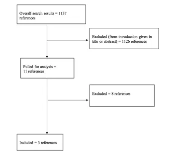
Levels of Evidence for Primary Research Questiona
| Types of studies | ||||
| Therapeutic studies – Investigating the results of treatment | Prognostic studies – Investigating the effect of a patient characteristic on the outcome of disease | Diagnostic studies – Investigating a diagnostic test | Economic and decision analyses – Developing an economic or decision model | |
| Level I | High-quality randomized trial with statistically significant difference or no statistically significant difference but narrow confidenceintervals Systematic reviewb of level I RCTs (and study results were homogenousc) | High-quality prospective studyd (all patients were enrolled at the same point in their disease with ≥80% follow-up of enrolled patients) Systematic reviewb of level I studies | Testing of previously developed diagnostic criteria on consecutive patients (with universally applied reference “gold” standard) Systematic reviewb of level I studies | Sensible costs and alternatives; values obtained from many studies; with multiway sensitivity analyses Systematic reviewb of level I studies |
| Level II | Lesser quality RCT (e.g., ≤80% follow-up, no blinding, or improper randomization) Prospectived comparative studye Systematic reviewb of level II studies or level I studies with inconsistent results | Retrospectivef study Untreated controls from an RCT Lesser quality prospective study (e.g., patients enrolled at different points in their disease or ≤80% follow-up) Systematic reviewb of level II studies | Development of diagnostic criteria on consecutive patients (with universally applied reference “gold” standard) Systematic reviewb of level II studies | Sensible costs and alternatives; values obtained from limited studies; with multiway sensitivity analyses Systematic reviewb of level II studies |
| Level III | Case control studyg Retrospectivef comparative studye Systematic reviewb of level III studies | Case control studyg | Study of non consecutive patients; without consistently applied reference “gold” standard Systematic reviewb of level III studies | Analyses based on limited alternatives and costs; and poor estimates Systematic reviewb of level III studies |
| Level IV | Case seriesh | Case series | Case-control study Poor reference standard | Analyses with no sensitivity analyses |
RCT, Randomized controlled trial.
aA complete assessment of quality of individual studies requires critical appraisal of all aspects of the study design.
bA combination of results from ≥2 previous studies.
cStudies provided consistent results.
dStudy was started before the first patient enrolled.
ePatients treated one way (e.g., instrumented arthrodesis) compared with a group of patients treated in another way (e.g., unsintrumented arthrodesis) at the same institution.
fThe study was started after the first patient enrolled.
gPatients identified for the study based on their outcome, called “cases” (e.g., pseudoarthrosis) are compared to those who did not have outcome, called “controls” (e.g., successful fusion).
hPatients treated one way with no comparison group of patients treated in another way.
Appendix IV. Linking Levels of Evidence to Grades of Recommendation
| Grade of Recommendation | Standard Language | Levels of Evidence | |
| A | Recommended | Two or more consistent level I studies | |
| B | Suggested | One level I study with additional supporting level II or III studies | Two or more consistent level II or III studies |
| C | Is an option | One level I, II, or III study with supporting level IV studies | Two or more consistent level IV studies |
| Insufficient (insufficient or conflicting evidence) | Insufficient evidence to make recommendation for or against | A single level I, II, III, or IV study without other supporting evidence | >1 study with inconsistent findingsa |
aNote that in the presence of multiple consistent studies, and a single outlying, inconsistent study, the Grade of Recommendation will be based on the level of the consistent studies.
Appendix V. Criteria Grading the Evidence
The task force used the criteria provided below to identify the strengths and weaknesses of the studies included in this guideline. Studies containing deficiencies were downgraded one level (no further downgrading allowed, unless so severe that study had to be excluded). Studies with no deficiencies based on study design and contained clinical information that dramatically altered current medical perceptions of topic were upgraded.
- Baseline study design (i.e., therapeutic, diagnostic, prognostic) determined to assign initial level of evidence.
- Therapeutic studies reviewed for following deficiencies:
- Failure to provide a power calculation for an RCT;
- High degree of variance or heterogeneity in patient populations with respect to presenting diagnosis/demographics or treatments applied;<80% of patient follow-up;Failure to utilize validated outcomes instrument;No statistical analysis of results;Cross over rate between treatment groups of >20%;Inadequate reporting of baseline demographic data;Small patient cohorts (relative to observed effects);Failure to describe method of randomization;Failure to provide flowchart following patients through course of study (RCT);Failure to account for patients lost to follow-up;Lack of independent post-treatment assessment (e.g., clinical, fusion status, etc.);Utilization of inferior control group:
- Historical controls;Simultaneous application of intervention and control within same patient.
- High degree of variance or heterogeneity in patient populations with respect to presenting diagnosis/demographics or treatments applied;<80% of patient follow-up;Failure to utilize validated outcomes instrument;No statistical analysis of results;Cross over rate between treatment groups of >20%;Inadequate reporting of baseline demographic data;Small patient cohorts (relative to observed effects);Failure to describe method of randomization;Failure to provide flowchart following patients through course of study (RCT);Failure to account for patients lost to follow-up;Lack of independent post-treatment assessment (e.g., clinical, fusion status, etc.);Utilization of inferior control group:
- Failure to provide a power calculation for an RCT;
- Methodology of diagnostic studies reviewed for following deficiencies:
- Failure to determine specificity and sensitivity;
- Failure to determine inter- and intraobserver reliability;
- Failure to provide correlation coefficient in the form of kappa values.
- Methodology of prognostic studies reviewed for following deficiencies:
- High degree of variance or heterogeneity in patient populations with respect to presenting diagnosis/demographics or treatments applied;
- Failure to appropriately define and assess independent and dependent variables (e.g., failure to use validated outcome measures when available).
Appendix VI. Evidence Tables
| Author, Year | Level of Evidence | Task Force Conclusions relative to question and rationale for evidence grading |
|---|---|---|
| Bailey et al,40 2014 | I | This paper provides evidence that nonoperative management without a brace is equivalent to treatment with a thoracolumbosacral orthosis for neurologically intact patients with AO type A3 burst fractures from T10–L3 |
| Post et al,42 2006 | IV | This paper provides evidence that the use of a brace does not improve functional outcome, disability, or pain compared to no brace in the nonoperative treatment of neurologically intact patients with type A thoracolumbar fractures |
| Shamji et al,41 2014 | II | This paper provides evidence that nonoperative treatment with or without a brace results in similar radiographic and clinical outcomes at 6 months in neurologically intact patients with thoracolumbar burst fracture. Treatment without brace results in a shorter length of hospitalization than treatment with a brace |
© Congress of Neurological Surgeons
Source: Neurosurgery, September 6, 2018
9. Operative Versus Nonoperative Treatment
Sponsored by: Congress of Neurological Surgeons and the Section on Disorders of the Spine and Peripheral Nerves in collaboration with the Section on Neurotrauma and Critical Care
Endorsed by: The Congress of Neurological Surgeons (CNS) and the American Association of Neurological Surgeons (AANS)
Craig H. Rabb, MD,1 Daniel J. Hoh, MD,2 Paul A. Anderson, MD,3 Paul M. Arnold, MD,4 John H. Chi, MD, MPH,5 Andrew T. Dailey, MD,6 Sanjay S. Dhall, MD,7 Kurt M. Eichholz, MD,8 James S. Harrop, MD,9 Sheeraz Qureshi, MD, MBA,10 P. B. Raksin, MD,11 Michael G. Kaiser, MD,12 and John E. O’Toole, MD, MS13
1. Department of Neurosurgery, University of Utah, Salt Lake City, Utah
2. Lillian S. Wells Department of Neurological Surgery, University of Florida, Gainesville, Florida
3. Department of Orthopedics and Rehabilitation, University of Wisconsin, Madison, Wisconsin
4. Department of Neurosurgery, University of Kansas School of Medicine, Kansas City, Kansas
5. Department of Neurosurgery, Harvard Medical School, Brigham and Women’s Hospital, Boston, Massachusetts
6. Department of Neurosurgery, University of Utah, Salt Lake City, Utah
7. Department of Neurological Surgery, University of California, San Francisco, San Francisco, California
8. St. Louis Minimally Invasive Spine Center, St. Louis, Missouri
9. Departments of Neurological Surgery and Orthopedic Surgery, Thomas Jefferson University, Philadelphia, Pennsylvania
10. Department of Orthopaedic Surgery, Weill Cornell Medical College, New York, New York
11. Division of Neurosurgery, John H. Stroger, Jr. Hospital of Cook County and Department of Neurological Surgery, Rush University Medical Center, Chicago, Illinois
12. Department of Neurosurgery, Columbia University, New York, New York
13. Department of Neurological Surgery, Rush University Medical Center, Chicago, Illinois
Correspondence:
Craig H. Rabb, MD
Department of Neurosurgery
University of Utah
175 North Medical Drive East
5th Floor, Neurosurgery
Salt Lake City, Utah, 84132
Email: craig.rabb@hsc.utah.edu
ABSTRACT
Background: There is a lack of consensus regarding the approach to treatment of a neurologically intact patient with a thoracic or lumbar burst fracture. Similarly, a widely adopted treatment paradigm for nonburst fractures, a diverse group, remains elusive.
Objective: We sought to determine the optimal treatment protocol for burst and nonburst fractures of the thoracic and lumbar spine.
Methods: A systematic review of the literature was performed using the National Library of Medicine PubMed database and the Cochrane Library for studies relevant to thoracolumbar fractures, including burst fractures and nonburst fractures. Clinical studies specifically comparing nonsurgical to surgical treatment for neurologically intact patients harboring burst and nonburst fractures were selected for review.
Results: The literature search yielded 836 abstracts. The task force selected 144 articles for full-text review, and 6 were selected for inclusion in this systematic review. Of these, 3 articles were level II and specifically studied burst fractures in neurologically intact patients. One article indicated the superiority of surgery, and 2 articles showed equivalence. There were 3 level III studies that provided conflicting evidence. There were no studies meeting the inclusion criteria that studied nonburst fractures.
Conclusion: With regard to patient outcomes, there is conflicting evidence for and against surgery in the management of neurologically intact patients with thoracolumbar burst fractures. Therefore, the task force recommends (grade insufficient) that the discretion of the treating provider should be used to determine if the presenting thoracic or lumbar burst fracture in the neurologically intact patient warrants surgical intervention. Similarly, because of the lack of studies specifically investigating nonburst fractures, this guideline provides a grade insufficient recommendation that the discretion of the treating provider should be used to determine if the presenting nonburst thoracolumbar fracture warrants surgical intervention.
RECOMMENDATIONS
| Question 1 |
| Does the surgical treatment of burst fractures of the thoracic and lumbar spine improve clinical outcomes compared to nonoperative treatment? |
| Recommendation 1 |
| There is conflicting evidence to recommend for or against the use of surgical intervention to improve clinical outcomes in patients with thoracolumbar burst fracture who are neurologically intact. Therefore, it is recommended that the discretion of the treating provider be used to determine if the presenting thoracic or lumbar burst fracture in the neurologically intact patient warrants surgical intervention. |
| Strength of Recommendation: Grade Insufficient |
| Question 2 |
| Does the surgical treatment of nonburst fractures of the thoracic and lumbar spine improve clinical outcomes compared to nonoperative treatment? |
| Recommendation 2 |
| There is insufficient evidence to recommend for or against the use of surgical intervention for nonburst thoracic or lumbar fractures. It is recommended that the decision to pursue surgery for such fractures be at the discretion of the treating physician. |
| Strength of Recommendation: Grade Insufficient |
INTRODUCTION
Goals and Rationale
This clinical guideline was created to improve patient care by outlining the appropriate information gathering and decision-making processes involved in the evaluation and treatment of patients with thoracolumbar spine trauma. The surgical management of these patients often takes place under a variety of circumstances and by various clinicians. This guideline was created as an educational tool to guide qualified physicians through a series of diagnostic and treatment decisions in an effort to improve the quality and efficiency of care.
The decision as to whether or not neurologically intact patients with thoracolumbar fractures require surgical intervention remains controversial. A consensus regarding the treatment of burst fractures, in particular, has been difficult to obtain. With the advent of modern spinal instrumentation, the options for surgical intervention have been refined considerably. The evolution of imaging techniques, such as magnetic resonance imaging (MRI) and reformatted computed tomography (CT) scans, has led to a better understanding of these injuries.
The most concerning complication related to nonoperative treatment of a patient with thoracolumbar fractures has been neurologic deterioration due to a failure to surgically decompress and/or stabilize the injured spine.1 More recently, physicians electing nonoperative care for neurologically intact patients are recognizing the potential for the progressive development of chronic pain and deformity. By contrast, surgeons should strive to determine the best treatment option for each individual patient, so as to avoid unnecessary surgery.
Historical treatment for thoracic and lumbar spine injuries has often been nonsurgical.2,3 Early discussions as to which fractures require surgical stabilization centered over the question of whether or not the injury was stable or unstable.4 White and Panjabi define clinical instability as “the loss of the ability of spine under physiologic loads to maintain relationships between vertebra in such a way that there is neither damage nor subsequent irritation to the spinal cord or nerve roots and, in addition, there is no development of incapacitating deformity or pain due to structural changes.” In determining which fractures require open stabilization, they were early advocates of determining whether the anterior and/or posterior elements were destroyed or unable to function. Denis5 later proposed via a case series further subdividing the anterior column by introducing the concept of a middle column, which included the posterior portion of the vertebral body, the posterior annulus, and posterior longitudinal ligament. The primary implication discussed was that posterior column injury is insufficient alone to cause instability. In 1998, Hitchon et al6 were among the first to propose that surgery was best reserved for patients with >20° of kyphosis, >50% height loss, or >50% canal stenosis. This study contained highly heterogeneous patient groups, including both intact patients and patients with spinal cord injuries. Moreover, diverse injury types were also included.
Over the past 20 years, the rapid evolution of both diagnostic imaging and spinal stabilization techniques has complicated interpretation of the literature, such that adequate determination of which patients would benefit most from surgical stabilization and/or decompression is difficult. For example, a number of early papers were based upon the use of Harrington instrumentation,7–10 which makes contemporary application of their findings challenging. Moreover, while few will argue that decompression and stabilization is appropriate for patients with neurologic deficits, the heterogeneity of patient samples, e.g., the presence or absence of neurologic deficits, has further complicated analysis of the literature.6 In addition, many older studies were performed before the widespread use of MRI, which may alter treatment decisions. A comprehensive assessment of the published literature devoted to this subject is critical to assist clinicians with decision-making as to which injuries require operative versus nonoperative treatment.
Methods
The guidelines task force initiated a systematic review of the literature relevant to the diagnosis and treatment of patients with thoracolumbar trauma. Through objective evaluation of the evidence and transparency in the process of making recommendations, this evidence-based clinical practice guideline was developed for the diagnosis and treatment of adult patients with thoracolumbar injury. These guidelines are developed for educational purposes to assist practitioners in their clinical decision-making processes. Additional information about the methods used in this systematic review can be found in the introduction and methodology chapter.
Literature Search
The task force members identified search terms/parameters, and a medical librarian implemented the literature search, consistent with the literature search protocol (see Appendix I), using the National Library of Medicine PubMed database and the Cochrane Library (which included the Cochrane Database of Systematic Reviews, the Database of Abstracts of Reviews of Effect, the Cochrane Central Register of Controlled Trials, the Health Technology Assessment Database, and the National Health Service Economic Evaluation Database) for the period from January 1, 1946 to March 31, 2015, using the search strategies provided in Appendix I.
RESULTS
The literature search yielded 836 abstracts. Task force members reviewed all abstracts yielded from the literature search and identified the literature for full-text review and extraction, addressing the clinical questions in accordance with the literature search protocol (Appendix I). Task force members identified the best research evidence available to answer the targeted clinical questions. When level I, II, and/or III literature was available to answer specific questions, the task force did not review level IV studies.
The task force selected 144 articles for full-text review. Of these, 138 were rejected for not meeting inclusion criteria or for being off- topic. Six were selected for inclusion in the systematic review (Appendix II).
Inclusion/Exclusion Criteria
Articles were retrieved and included only if they met specific inclusion/exclusion criteria. These criteria were also applied to articles provided by guideline task force members who supplemented the electronic database searches with articles from their own files. To reduce bias, these criteria were specified before conducting the literature searches.
Articles that do not meet the following criteria were, for the purposes of this evidence-based clinical practice guideline, excluded. To be included as evidence in the guideline, an article had to be a report of a study that:
- Investigated patients with thoracolumbar injuries;
- Included patients ≥18 years of age;
- Enrolled ≥80% of thoracolumbar injuries (studies with mixed patient populations were included if they reported results separately for each group/patient population);
- Was a full article report of a clinical study;
- Was not an internal medical records review, meeting abstract, historical article, editorial, letter, or commentary;
- Appeared in a peer-reviewed publication or a registry report;
- Enrolled ≥10 patients per arm per intervention (20 total) for each outcome;
- Included only human subjects;
- Was published in or after 1946 through March 31, 2015;
- Quantitatively presented results;
- Was not an in vitro study;
- Was not a biomechanical study;
- Was not performed on cadavers;
- Was published in English;
- Was not a systematic review, meta-analysis, or guideline developed by others*:
Rating Quality of Evidence
The guideline task force used a modified version of the North American Spine Society’s evidence-based guideline development methodology. The North American Spine Society methodology uses standardized levels of evidence (Appendix III) and grades of recommendation (Appendix IV) to assist practitioners in easily understanding the strength of the evidence and the recommendations within the guidelines. The levels of evidence range from level I (high-quality randomized controlled trial) to level IV (case series). Grades of recommendation indicate the strength of the recommendations made in the guideline based on the quality of the literature. Levels of evidence have specific criteria and are assigned to studies prior to developing recommendations. Recommendations are then graded based upon the level of evidence. To better understand how levels of evidence inform the grades of recommendation and the standard nomenclature used within the recommendations, see Appendix IV.
Guideline recommendations were written using a standard language that indicates the strength of the recommendation. “A” recommendations indicate a test or intervention is “recommended”; “B” recommendations “suggest” a test or intervention; “C” recommendations indicate a test or intervention or “is an option.” “Insufficient evidence” statements clearly indicate that “there is insufficient evidence to make a recommendation for or against” a test or intervention. Task force consensus statements clearly state that “in the absence of reliable evidence, it is the task force’s opinion that” a test or intervention may be considered. Both the levels of evidence assigned to each study and the grades of each recommendation were arrived at by consensus of the workgroup employing up to three rounds of voting when necessary.
In evaluating studies as to levels of evidence for this guideline, the study design was interpreted as establishing only a potential level of evidence. For example, a therapeutic study designed as a randomized controlled trial would be considered a potential level I study. The study would then be further analyzed as to how well the study design was implemented, and significant shortcomings in the execution of the study would be used to downgrade the levels of evidence for the study’s conclusions (see Appendix V for additional information and criteria).
Revision Plans
In accordance with the Institute of Medicine’s standards for developing clinical practice guidelines and criteria specified by the National Guideline Clearinghouse, the task force will monitor related publications following the release of this document and will revise the entire document and/or specific sections “if new evidence shows that a recommended intervention causes previously unknown substantial harm; that a new intervention is significantly superior to a previously recommended intervention from an efficacy or harms perspective; or that a recommendation can be applied to new populations.”11 In addition, the task force will confirm within 5 years from the date of publication that the content reflects current clinical practice and the available technologies for the evaluation and treatment for patients with thoracolumbar trauma.
DISCUSSION
| Question 1 |
| Does the surgical treatment of burst fractures of the thoracic and lumbar spine improve clinical outcomes compared to nonoperative treatment? |
| Recommendation 1 |
| There is conflicting evidence to recommend for or against the use of surgical intervention to improve clinical outcomes in patients with thoracolumbar burst fractures who are neurologically intact. Therefore, it is recommended that the discretion of the treating provider be used to determine if the presenting thoracic or lumbar burst fracture in the neurologically intact patient warrants surgical intervention. |
| Strength of Recommendation: Grade Insufficient |
Level II Evidence
None of the studies met the criteria to be considered level I evidence. There were 3 class II studies.8–10 While these studies were randomized controlled trials (RCTs), various flaws led to downgrading them to level II evidence. In the study by Shen et al12 outcomes were similar at 2 years, when comparing nonoperative treatment to short-segment posterior pedicle screw fixation. Siebenga et al13 also compared operative versus nonoperative management; however, this study suffers from a small sample size (34 patients). The authors looked at patients with AO type A fractures who were neurologically intact and, by analyzing subgroups, they concluded that patients with AO type A3 (burst) fractures fare better with short-segment posterior fixation, in terms of both radiographic and functional assessment. In the surgically treated group, more patients returned to work than in the nonoperatively treated group. The study by Wood et al,14 although considered an RCT, fell short of being considered level I evidence. The authors specifically excluded cases thought to have posterior ligamentous disruption. In addition, <80% of the patients had adequate follow-up. There were 24 patients treated operatively and 23 treated nonoperatively. No significant differences were found regarding return to work, pain scores, or kyphosis. The authors were unable to confirm their hypothesis that surgery (performed via a posterior approach) was superior to nonoperative treatment.
Level III Evidence
Some comparative studies met inclusion criteria but were downgraded to level III. Landi et al15 performed a retrospective comparative study (25 patients in each arm). Follow-up was conducted at 3 and 6 months, comparing percutaneous screw fixation to bracing for Magerl type A3 fractures. Patients had better satisfaction with surgery. Another study included patients with A1 and A2 fractures (including nonburst fractures) and concluded that outcomes with surgery were superior.16
Wood conducted a long-term follow-up study of patients previously studied in 2003.14,17 This study included a small number of patients, who were consecutively assigned and randomized to operative treatment or nonoperative treatment, but the method of randomization was not reported. Therefore, the study was downgraded to level II. There was also a lack of blinding, small cohorts, and no power analysis was performed. The study was further downgraded to level III, showing an advantage to nonoperative care over surgery.
| Question 2 |
| Does the surgical treatment of nonburst fractures of the thoracic and lumbar spine improve clinical outcomes compared to nonoperative treatment? |
| Recommendation 2 |
| There is insufficient evidence to recommend for or against the use of surgical intervention for nonburst thoracolumbar fractures. It is recommended that the decision to pursue surgery for such fractures be at the discretion of the treating physician. |
| Strength of Recommendation: Grade Insufficient |
Level IV Evidence
None of the studies examined met the criteria for inclusion. The literature search located a number of retrospective comparative studies of surgical versus nonsurgical treatment of thoracic and lumbar fractures.18–20 The studies included patients with varying degrees of neurologic deficits and/or instability. In addition, fractures that the authors considered “unstable” underwent surgery. These patients were not consistently classified by any standard manner to deem them “unstable,” thereby calling into question the possible heterogeneity of the comparison groups. One study of patients presenting with paraplegia compared nonoperative treatment to laminectomy with or without fusion and to patients receiving Harrington instrumentation. The authors concluded that laminectomy was inferior to either Harrington instrumentation or conservative treatment, and that pain complaints were worse in the laminectomy group and in the conservatively treated group.21 An additional key issue that caused these papers to be downgraded is that many older retrospective comparative studies involved patients who underwent surgical procedures using outdated instrumentation (e.g., Harrington rod fixation) and noncurrent diagnostic imaging (e.g., plain x-ray). Therefore, the relevance of these studies in the modern era is questionable. As a result, unfortunately, adequate studies in this area are currently lacking.
Future Research
As this literature review has demonstrated, there is a need for further research regarding operative versus nonoperative treatment of patients with burst or nonburst thoracolumbar fractures. With respect to burst fractures, given the rapid evolution of imaging, a focus on the posterior ligamentous complex (PLC) in neurologically intact patients should be more thoroughly investigated. An RCT of neurologically intact patients with and without disruption of the PLC may help to more definitively answer this question.
Heterogeneity of thoracolumbar injuries has hindered the interpretation of the literature with regard to nonburst fractures, as no high-quality randomized controlled trials exist in this area. It may prove to be too challenging ethically to try to perform such studies. A trial specifically dedicated to classic bony Chance fractures, for example, could shed some light on this subject, but in all probability, it may not be feasible. Likewise, comparing the likelihood of developing posttraumatic cord tethering and/or syringomyelia in spinal cord injury patients who received either operative or nonoperative treatment would be of interest. It may prove that prospective registries of patients treated for various nonburst thoracolumbar fractures provide the greatest amount of information to guide treatment decisions.
Conclusions
Most surgeons today use surgical intervention for patients with thoracolumbar fractures who present with neurologic deficits, owing to assumed instability, and the desire to restore alignment, decompress neural elements, and stabilize the spine to reduce pain, prevent deformity, and allow for early mobilization. There is little research available for the neurologically intact patient. Relatively high-quality studies have been performed for patients with burst fractures, but have yielded conflicting conclusions, such that either surgery or nonoperative treatment remain viable options. Unfortunately, high-quality studies have yet to be performed to investigate which option results in the best outcomes for nonburst fractures. As such, it must be left to the discretion of the treating surgeon as to which treatment option is best for a given patient.
Potential Conflicts of Interest
The task force members were required to report all possible conflicts of interest (COIs) prior to beginning work on the guideline, using the COI disclosure form of the AANS/CNS Joint Guidelines Review Committee, including potential COIs that are unrelated to the topic of the guideline. The CNS Guidelines Committee and Guideline Task Force Chairs reviewed the disclosures and either approved or disapproved the nomination. The CNS Guidelines Committee and Guideline Task Force Chairs are given latitude to approve nominations of Task Force members with possible conflicts and address this by restricting the writing and reviewing privileges of that person to topics unrelated to the possible COIs. The conflict of interest findings are provided in detail in the companion introduction and methods manuscript.
Disclaimer of Liability
This clinical systematic review and evidence-based guideline was developed by a multidisciplinary physician volunteer task force and serves as an educational tool designed to provide an accurate review of the subject matter covered. These guidelines are disseminated with the understanding that the recommendations by the authors and consultants who have collaborated in their development are not meant to replace the individualized care and treatment advice from a patient’s physician(s). If medical advice or assistance is required, the services of a competent physician should be sought. The proposals contained in these guidelines may not be suitable for use in all circumstances. The choice to implement any particular recommendation contained in these guidelines must be made by a managing physician in light of the situation in each particular patient and on the basis of existing resources.
Disclosures
These evidence-based clinical practice guidelines were funded exclusively by the Congress of Neurological Surgeons and the Section on Disorders of the Spine and Peripheral Nerves in collaboration with the Section on Neurotrauma and Critical Care, which received no funding from outside commercial sources to support the development of this document.
Acknowledgments
The guidelines task force would like to acknowledge the CNS Guidelines Committee for their contributions throughout the development of the guideline and the AANS/CNS Joint Guidelines Review Committee for their review, comments, and suggestions throughout peer review, as well as the contributions of Trish Rehring, MPH, CHES, Senior Manager of Clinical Practice Guidelines for the CNS, and Mary Bodach, MLIS, Guidelines Specialist and Medical Librarian for assistance with the literature searches. Throughout the review process the reviewers and authors were blinded from one another. At this time, the guidelines task force would like to acknowledge the following individual peer reviewers for their contributions: Maya Babu, MD, MBA, Greg Hawryluk, MD, PhD, Steven Kalkanis, MD, Yi Lu, MD, PhD, Jeffrey J. Olson, MD, Martina Stippler, MD, Cheerag Upadhyaya, MD, MSc, and Robert Whitmore, MD.
REFERENCES
1. Denis F, Armstrong GW, Searls K, Matta L. Acute thoracolumbar burst fractures in the absence of neurologic deficit. A comparison between operative and nonoperative treatment. Clin Orthop Relat Res 1984;189:142-149.
2. Guttmann L. Spinal deformities in traumatic paraplegics and tetraplegics following surgical procedures. Paraplegia 1969;7:38-58.
3. Bedbrook GM. Treatment of thoracolumbar dislocation and fractures with paraplegia. Clin Orthop Relat Res 1975;112:27-43.
4. White AA, Panjabi MM. Clinical biomechanics of the spine. Philadelphia: Lippincott; 1978.
5. Denis F. The three column spine and its significance in the classification of acute thoracolumbar spinal injuries. Spine 1983;8:817-831.
6. Hitchon PW, Torner JC, Haddad SF, Follett KA. Management options in thoracolumbar burst fractures. Surg Neurol 1998;49:619-626.
7. Jacobs RR, Asher MA, Snider RK. Thoracolumbar spinal injuries. A comparative study of recumbent and operative treatment in 100 patients. Spine 1980;5:463-477.
8. Lifeso RM, Arabie KM, Kadhi SK. Fractures of the thoraco-lumbar spine. Paraplegia 1985;23:207-224.
9. Lindahl S, Willen J, Irstam L. Unstable thoracolumbar fractures. A comparative radiologic study of conservative treatment and Harrington instrumentation. Acta Radiol Diagn (Stockh) 1985;26:67-77.
10. McEvoy RD, Bradford DS. The management of burst fractures of the thoracic and lumbar spine. Experience in 53 patients. Spine 1985;10:631-637.
11. Ransohoff DF, Pignone M, Sox HC. How to decide whether a clinical practice guideline is trustworthy. JAMA 2013;309:139-140.
12. Shen WJ, Liu TJ, Shen YS. Nonoperative treatment versus posterior fixation for thoracolumbar junction burst fractures without neurologic deficit. Spine 2001;26:1038-1045.
13. Siebenga J, Leferink VJ, Segers MJ, et al. Treatment of traumatic thoracolumbar spine fractures: a multicenter prospective randomized study of operative versus nonsurgical treatment. Spine 2006;31:2881-2890.
14. Wood K, Buttermann G, Mehbod A, Garvey T, Jhanjee R, Sechriest V. Operative compared with nonoperative treatment of a thoracolumbar burst fracture without neurological deficit. A prospective, randomized study. J Bone Joint Surg Am 2003;85A:773-781.
15. Landi A, Marotta N, Mancarella C, Meluzio MC, Pietrantonio A, Delfini R. Percutaneous short fixation vs conservative treatment: comparative analysis of clinical and radiological outcome for A.3 burst fractures of thoraco-lumbar junction and lumbar spine. Eur Spine J 2014;23(suppl 6):671-676.
16. Medici A, Meccariello L, Falzarano G. Non-operative vs. percutaneous stabilization in Magerl’s A1 or A2 thoracolumbar spine fracture in adults: is it really advantageous for a good alignment of the spine? Preliminary data from a prospective study. Eur Spine 2014;23(suppl 6):677-683.
17. Wood KB, Buttermann GR, Phukan R, et al. Operative compared with nonoperative treatment of a thoracolumbar burst fracture without neurological deficit: a prospective randomized study with follow-up at sixteen to twenty-two years. J Bone Joint Surg Am 2015;97:3-9.
18. Fang D, Leong JC, Cheung HC. The treatment of thoracolumbar spinal injuries with paresis by conservative versus surgical methods. Ann Acad Med 1982;11:203-206.
19. Soreff J, Axdorph G, Bylund P, Odeen I, Olerud S. Treatment of patients with unstable fractures of the thoracic and lumbar spine: a follow-up study of surgical and conservative treatment. Acta Orthop Scand 1982;53:369-381.
20. Willén J, Lindahl S, Nordwall A. Unstable thoracolumbar fractures. A comparative clinical study of conservative treatment and Harrington instrumentation. Spine 1985;10:111-122.
21. Willén J, Dahllöf AG, Nordwall A. Paraplegia in unstable thoracolumbar injuries. A study of conservative and operative treatment regarding neurological improvement and rehabilitation. Scand J Rehabil Med Suppl 1983;9:195-205.
Appendix I. Literature Searches
Search Strategies
PubMed
- Lumbar vertebrae [MeSH] OR Thoracic vertebrae [MeSH]
- Spinal Injuries [MeSH] OR Spinal Cord Injuries [MeSH]
- #1 AND #2
- Thoracolumbar [TIAB] OR thoraco-lumbar [TIAB] OR thoraco lumbar [TIAB] OR burst [Title]
- Injur* [TIAB] OR trauma* [TIAB] OR fractur* [TIAB] OR dislocation* [TIAB]
- #4 AND #5
- Lumbar vertebrae/injuries [MeSH] OR Thoracic vertebrae/injuries [MeSH] (3150 results)
- #3 OR #6 OR #7
- Braces [MeSH] OR Casts, Surgical [MeSH] OR Bed rest [MeSH] OR Physical Therapy Modalities[MeSH] OR Rehabilitation [MeSH] OR rehabilitation[SH]
- Drug therapy[sh] OR Analgesics[mh] OR analgesics[pa] OR “Muscle Relaxants, Central”[mh] OR Steroids[mh] OR Glucocorticoids[mh] OR Glucocorticoids[pa]
- Brace OR braces OR bracing OR orthos* OR orthotic* OR cast OR casts OR casting OR TSLO [TIAB]
- Bed rest OR bedrest OR “physical therapy” OR physiotherap* OR rehabilitation [TIAB]
- NSAID[tiab] OR opioid*[tiab] OR (muscle[tiab] AND relax*[tiab]) OR acetaminophen[tiab] OR naproxen[tiab] OR ibuprofen[tiab] OR hydrocodone[tiab] OR oxycodone[tiab] OR oxycontin[tiab] OR morphine[tiab] OR benzodiazepine*[tiab] OR tramadol[tiab] OR steroid*[tiab] OR prednisone[tiab] OR solumedrol[tiab] OR fentanyl[tiab] OR lidoderm[tiab] OR aspirin[tiab] OR codeine[tiab] OR drug* [TIAB] OR medication* [TIAB]
- (Conservative[tiab] OR non-operat*[tiab] OR nonoperat*[tiab] OR non-surg*[tiab] OR nonsurg*[tiab]) AND (treatment*[tiab] OR therap*[tiab] OR management[tiab])
- #9 OR #10 OR #11 OR #12 OR #13 OR #14
- Orthopedic Procedures [MeSH] OR Neurosurgical Procedures [MeSH] OR Decompression, surgical [MeSH] OR Orthopedic Fixation Devices [MeSH] OR surgery [SH] OR instrumentation [SH]
- surgery[tiab] OR surgical[tiab] OR operati*[tiab] OR repair*[tiab] OR stabiliz*[tiab] OR fixation[tiab] OR reconstruct*[tiab] OR fusion[tiab] OR decompress*[tiab] OR spondylodes*[tiab] OR spondylosyndes*[tiab] OR arthrodes*[tiab] OR laminectomy[tiab] OR discectomy[tiab] OR diskectomy OR “percutaneous vertebral augmentation”[tiab] OR “bone screw”[tiab] OR “bone screws”[tiab] OR “bone plate”[tiab] OR “bone plates”[tiab] OR “pedicle screw”[tiab] OR “pedicle screws”[tiab]
- #16 OR #17
- #8 AND (#15 AND #18)
- (animal [MeSH] NOT human [MeSH]) OR cadaver [MeSH] OR cadaver* [Titl] OR comment [PT] OR letter [PT] OR editorial [PT] OR addresses [PT] OR news [PT] OR “newspaper article” [PT] OR case reports [PT]
- #19 NOT #20
- osteoporosis [MH] OR osteoporotic fractures [MH] OR osteoporo* [TITLE] OR spinal neoplasms [MH] OR tumor* [TITLE] OR tumour* [TITLE] OR malignan* [TITLE]
- #21 NOT #22
- #23 AND English [Lang]
Cochrane Library
- Lumbar vertebrae: MeSH descriptor, explode all trees
- Thoracic vertebrae: MeSH descriptor, explode all trees
- #1 OR #2
- Spinal Injuries: MeSH descriptor
- Spinal Cord Injuries: MeSH descriptor
- #4 OR #5
- #3 AND #6
- (Thoracolumbar OR thoraco-lumbar OR thoraco lumbar OR burst) NEAR/4 (Injur* OR trauma* OR fractur* OR dislocation*):ti,ab,kw
- Lumbar vertebrae/injuries: MeSH descriptor, explode all trees
- Thoracic vertebrae/injuries: MeSH descriptor, explode all trees
- #9 OR #10
- #7 OR #8 OR #11
- mh osteoporosis or mh osteoporotic fractures or mh spinal neoplasms
- osteoporo* or tumor* or malignan*:ti
- #13 OR #14
- #12 NOT #15
Appendix II. Article Inclusions and Exclusions
Included and Excluded Articles Flowchart

Appendix III. Rating Evidence Quality
Levels of Evidence for Primary Research Questiona
| Types of studies | ||||
| Therapeutic studies – Investigating the results of treatment | Prognostic studies – Investigating the effect of a patient characteristic on the outcome of disease | Diagnostic studies – Investigating a diagnostic test | Economic and decision analyses – Developing an economic or decision model | |
| Level I | High-quality randomized trial with statistically significant difference or no statistically significant difference but narrow confidenceintervals Systematic reviewb of level I RCTs (and study results were homogenousc) | High-quality prospective studyd (all patients were enrolled at the same point in their disease with ≥80% follow-up of enrolled patients) Systematic reviewb of level I studies | Testing of previously developed diagnostic criteria on consecutive patients (with universally applied reference “gold” standard) Systematic reviewb of level I studies | Sensible costs and alternatives; values obtained from many studies; with multiway sensitivity analyses Systematic reviewb of level I studies |
| Level II | Lesser quality RCT (e.g., ≤80% follow-up, no blinding, or improper randomization) Prospectived comparative studye Systematic reviewb of level II studies or level I studies with inconsistent results | Retrospectivef study Untreated controls from an RCT Lesser quality prospective study (e.g., patients enrolled at different points in their disease or ≤80% follow-up) Systematic reviewb of level II studies | Development of diagnostic criteria on consecutive patients (with universally applied reference “gold” standard) Systematic reviewb of level II studies | Sensible costs and alternatives; values obtained from limited studies; with multiway sensitivity analyses Systematic reviewb of level II studies |
| Level III | Case control studyg Retrospectivef comparative studye Systematic reviewb of level III studies | Case control studyg | Study of non consecutive patients; without consistently applied reference “gold” standard Systematic reviewb of level III studies | Analyses based on limited alternatives and costs; and poor estimates Systematic reviewb of level III studies |
| Level IV | Case seriesh | Case series | Case-control study Poor reference standard | Analyses with no sensitivity analyses |
RCT, Randomized controlled trial.
aA complete assessment of quality of individual studies requires critical appraisal of all aspects of the study design.
bA combination of results from ≥2 previous studies.
cStudies provided consistent results.
dStudy was started before the first patient enrolled.
ePatients treated one way (e.g., instrumented arthrodesis) compared with a group of patients treated in another way (e.g., unsintrumented arthrodesis) at the same institution.
fThe study was started after the first patient enrolled.
gPatients identified for the study based on their outcome, called “cases” (e.g., pseudoarthrosis) are compared to those who did not have outcome, called “controls” (e.g., successful fusion).
hPatients treated one way with no comparison group of patients treated in another way.
Appendix IV. Linking Levels of Evidence to Grades of Recommendation
| Grade of Recommendation | Standard Language | Levels of Evidence | |
| A | Recommended | Two or more consistent level I studies | |
| B | Suggested | One level I study with additional supporting level II or III studies | Two or more consistent level II or III studies |
| C | Is an option | One level I, II, or III study with supporting level IV studies | Two or more consistent level IV studies |
| Insufficient (insufficient or conflicting evidence) | Insufficient evidence to make recommendation for or against | A single level I, II, III, or IV study without other supporting evidence | >1 study with inconsistent findingsa |
aNote that in the presence of multiple consistent studies, and a single outlying, inconsistent study, the Grade of Recommendation will be based on the level of the consistent studies.
Appendix V. Criteria Grading the Evidence
The task force used the criteria provided below to identify the strengths and weaknesses of the studies included in this guideline. Studies containing deficiencies were downgraded one level (no further downgrading allowed, unless so severe that study had to be excluded). Studies with no deficiencies based on study design and contained clinical information that dramatically altered current medical perceptions of topic were upgraded.
- Baseline study design (i.e., therapeutic, diagnostic, prognostic) determined to assign initial level of evidence.
- Therapeutic studies reviewed for following deficiencies:
- Failure to provide a power calculation for an RCT;
- High degree of variance or heterogeneity in patient populations with respect to presenting diagnosis/demographics or treatments applied;
- <80% of patient follow-up;
- Failure to utilize validated outcomes instrument;
- No statistical analysis of results;
- Cross over rate between treatment groups of >20%;
- Inadequate reporting of baseline demographic data;
- Small patient cohorts (relative to observed effects);
- Failure to describe method of randomization;
- Failure to provide flowchart following patients through course of study (RCT);
- Failure to account for patients lost to follow-up;
- Lack of independent post-treatment assessment (e.g., clinical, fusion status, etc.);
- Utilization of inferior control group:
- Historical controls;
- Simultaneous application of intervention and control within same patient.
- Failure to standardize surgical/intervention technique;
- Inadequate radiographic technique to determine fusion status (e.g., static radiographs for instrumented fusion).
- Methodology of diagnostic studies reviewed for following deficiencies:
- Failure to determine specificity and sensitivity;
- Failure to determine inter- and intraobserver reliability;
- Failure to provide correlation coefficient in the form of kappa values.
- Methodology of prognostic studies reviewed for following deficiencies:
- High degree of variance or heterogeneity in patient populations with respect to presenting diagnosis/demographics or treatments applied;
- Failure to appropriately define and assess independent and dependent variables (e.g., failure to use validated outcome measures when available).
Appendix VI. Evidence Tables
| Author, Year | Level of Evidence | Task Force Conclusions Relative to Question and Rationale for Evidence Grading |
| Landi et al,15 2014 | III | This paper provides evidence that patients had better satisfaction with percutaneous screw fixation surgery. Retrospective comparative study (25 patients in each arm). Follow-up was conducted at 3 and 6 months, comparing percutaneous screw fixation to bracing for Magerl type A3 fractures |
| Medici et al,16 2014 | III | This paper provides evidence that patients who had percutaneous fixation had better functional outcomes than patients treated nonoperatively with a 3-point orthopedic corset. This study included patients with A1 andA2 fractures (including nonburst). This study is a level III study; authors are assuming retrospective. This is the only study that includes nonburst fractures |
| Shen et al,12 2001 | II | This paper provides evidence that short-segment posterior fixation provides partial kyphosis correction and earlier pain relief, but the functional outcome at 2 years is similar. This is a prospective study, not an RCT. This study was assigned a level II, with equivalent outcomes |
| Siebenga et al,13 2006 | II | This paper provides evidence that patients with a type A3 thoracolumbar spine fracture without neurologic deficit should be treated by short-segment posterior stabilization. This study included 34 patients and concluded the opposite of what has been shown previously, with longer follow-up (4-year follow-up), but at a lower rate. This is an RCT but was downgraded from level I to II because of the small number of patients |
| Wood et al,14 2003 | II | This paper provides evidence that operative treatment of patients with a stable thoracolumbar burst fracture and normal findings on the neurological examination provided no major long-term advantage compared with nonoperative treatment. This study was downgraded from level I to II, showing no difference between operative vs. nonoperative patients |
| Wood et al,17 2015 | III | This paper provides evidence that while early analysis (4 years) revealed few significant differences between the 2 groups, at long-term follow-up (16–22 years), those patients with a stable burst fracture who were treated nonoperatively reported less pain and better function compared with those who were treated surgically. This study is an RCT, but guideline authors question if the loss of more patients to follow-up can skew the data. It includes a small number of patients, who were consecutively assigned and randomized to operative treatment or nonoperative treatment, but the method of randomization was not reported. Therefore, the study should be downgraded from level I to level II. There was also a lack of blinding, small cohorts, and no power analysis was performed. Therefore, the study was further downgraded to level III, showing an advantage to nonoperative care |
RCT = randomized controlled trial.
© Congress of Neurological Surgeons
Source: Neurosurgery, September 6, 2018
10. Timing of Surgical Intervention
Sponsored by: Congress of Neurological Surgeons and the Section on Disorders of the Spine and Peripheral Nerves in collaboration with the Section on Neurotrauma and Critical Care
Endorsed by: The Congress of Neurological Surgeons (CNS) and the American Association of Neurological Surgeons (AANS)
Kurt M. Eichholz, MD,1 Craig H. Rabb, MD,2 Paul A. Anderson, MD,3 Paul M. Arnold, MD,4 John H. Chi, MD, MPH,5 Andrew T. Dailey, MD,6 Sanjay S. Dhall, MD,7 James S. Harrop, MD,8 Daniel J. Hoh, MD,9 Sheeraz Qureshi, MD, MBA,10 P. B. Raksin, MD,11 Michael G. Kaiser, MD,12 and John E. O’Toole, MD, MS13
1. St. Louis Minimally Invasive Spine Center, St. Louis, Missouri
2. Department of Neurosurgery, University of Utah, Salt Lake City, Utah
3. Department of Orthopedics and Rehabilitation, University of Wisconsin, Madison, Wisconsin
4. Department of Neurosurgery, University of Kansas School of Medicine, Kansas City, Kansas
5. Department of Neurosurgery, Harvard Medical School, Brigham and Women’s Hospital, Boston, Massachusetts
6. Department of Neurosurgery, University of Utah, Salt Lake City, Utah
7. Department of Neurological Surgery, University of California, San Francisco, San Francisco, California
8. Departments of Neurological Surgery and Orthopedic Surgery, Thomas Jefferson University, Philadelphia, Pennsylvania
9. Lillian S. Wells Department of Neurological Surgery, University of Florida, Gainesville, Florida
10. Department of Orthopaedic Surgery, Weill Cornell Medical College, New York, New York
11. Division of Neurosurgery, John H. Stroger, Jr. Hospital of Cook County and Department of Neurological Surgery, Rush University Medical Center, Chicago, Illinois
12. Department of Neurosurgery, Columbia University, New York, New York
13. Department of Neurological Surgery, Rush University Medical Center, Chicago, Illinois
Correspondence:
Kurt M. Eichholz, MD
St. Louis Minimally Invasive Spine Center
4590 South Lindbergh Blvd.
St. Louis, MO 63126
Email: kurt@stlmisc.com
Keywords: Thoracic and lumbar fractures, thoracic and lumbar fusion, timing of surgery, treatment of thoracic and lumbar fractures
Abbreviations
ASIA – American Spinal Injury Association
ISS – Injury Severity Score
NASS – North American Spine Society
ABSTRACT
Background: Thoracolumbar fractures include complete and incomplete spinal cord injuries. Treatments for these fractures range from conservative therapy with bedrest and bracing, to decompression and stabilization of the spine with instrumentation. There are many variables that may affect the outcome of patients undergoing such treatments. The effect of the time between injury and surgical intervention has not been well established.
Objective: To determine if the timing of surgical intervention influences the neurological outcome for patients with thoracic and lumbar fractures.
Methods: The National Library of Medicine PubMed and the Cochrane Library databases were searched for publications related to the timing of surgical intervention and thoracic and lumbar fracture treatment. After exclusion criteria were applied, full-text articles were reviewed by the authors and the Task Force. Studies were graded based on a modified North American Spine Society evidence-based guideline development methodology, and recommendations were made by the Task Force based on the review of the full-text articles.
Results: A total of 1172 abstracts were screened. Of these, 69 full-text articles were reviewed. Fifty-eight were rejected for not meeting inclusion criteria. Eleven articles were selected for inclusion in this systematic review. Two studies provided level III evidence, while 9 studies provided level IV evidence. There was inconsistency in these studies regarding the effects of timing of surgical intervention on the neurological outcome of patients with thoracic and lumbar fractures, as well as inconsistency regarding what amount of time constituted “early” versus “late” surgical intervention.
Conclusion: There is insufficient and conflicting evidence regarding the effect of timing of surgical intervention on neurological outcomes in patients with thoracic and lumbar fractures. It is suggested that “early” surgery be considered as an option in patients with thoracic and lumbar fractures to reduce length of stay and complications. The available literature has defined “early” surgery inconsistently, ranging from <8 hours to <72 hours after injury.
RECOMMENDATIONS
| Question |
| Does early surgical intervention improve outcomes for patients with thoracic and lumbar fractures? |
| Recommendations |
| There is insufficient and conflicting evidence regarding the effect of timing of surgical intervention on neurological outcomes in patients with thoracic and lumbar fractures. |
| Strength of Recommendation: Grade Insufficient |
| It is suggested that “early” surgery be considered as an option in patients with thoracic and lumbar fractures to reduce length of stay and complications. The available literature has defined “early” surgery inconsistently, ranging from <8 hours to <72 hours after injury. |
| Strength of Recommendation: Grade B |
INTRODUCTION
Goals and Rationale
This clinical guideline has been created to improve patient care by outlining the appropriate information gathering and decision-making processes involved in the evaluation and treatment of patients with thoracolumbar spine trauma. The surgical management of these patients often takes place under a variety of circumstances and by various clinicians. This guideline has been created as an educational tool to guide qualified physicians through a series of diagnostic and treatment decisions to improve the quality and efficiency of care.
Background
Thoracolumbar fractures include a variety of traumatic spine injuries, both in isolation and in conjunction with other injuries. Over the decades, the treatment of thoracolumbar fractures has significantly evolved. Traditional conservative therapy involved bed rest and bracing. Adverse consequences of prolonged bed rest and the evolution of surgical technique have led to effective operative approaches for stabilization of thoracolumbar fractures. In many circumstances, surgery is now considered the optimal treatment for patients presenting with unstable thoracolumbar spine fractures.
However, the timing of surgical intervention in the setting of thoracolumbar fractures has been debated over the years, and the relationship of timing of surgical intervention to clinical outcome has not been well defined. While one might argue that “early” or “late” surgery may or may not improve clinical outcome, even the definition of what constitutes “early” versus “late” surgery is not well defined.
The goal of this guideline is to evaluate the available literature to determine if the timing of surgical intervention has an effect of the clinical outcome for patients suffering thoracic and lumbar fractures.
Methods
The guidelines task force initiated a systematic review of the literature relevant to the diagnosis and treatment of patients with thoracolumbar trauma. Through objective evaluation of the evidence and transparency in the process of making recommendations, this evidence-based clinical practice guideline was developed for the diagnosis and treatment of adult patients with thoracolumbar injury. These guidelines were developed for educational purposes to assist practitioners in their clinical decision-making processes. Additional information about the methods utilized in this systematic review can be found in the introduction and methodology chapter.
Literature Search
The task force members identified search terms/parameters, and a medical librarian implemented the literature search, consistent with the literature search protocol (see Appendix I), using the National Library of Medicine PubMed database and the Cochrane Library (which included the Cochrane Database of Systematic Reviews, the Database of Abstracts of Reviews of Effect, the Cochrane Central Register of Controlled Trials, the Health Technology Assessment Database, and the National Health Service Economic Evaluation Database) for the period from January 1, 1946, to March 31, 2015, using the search strategies provided in Appendix I.
RESULTS
The literature search yielded 1172 abstracts. Task force members reviewed all abstracts yielded from the literature search and identified the literature for full-text review and extraction, addressing the clinical questions, in accordance with the literature search protocol (Appendix I). Task force members identified the best research evidence available to answer the targeted clinical questions. When level I, II, and/or III literature was available to answer specific questions, the task force did not review level IV studies.
The task force selected 69 articles for full text review. Of these, 58 articles were rejected for not meeting inclusion criteria or for being off topic. Eleven studies were selected for inclusion in this systematic review (Appendix II).
Inclusion/Exclusion Criteria
Articles were retrieved and included only if they met specific inclusion/exclusion criteria. These criteria were also applied to articles provided by guideline task force members who supplemented the electronic database searches with articles from their own files. To reduce bias, these criteria were specified before conducting the literature searches.
Articles that do not meet the following criteria were, for the purposes of this evidence-based clinical practice guideline, were excluded. To be included as evidence in the guideline, an article had to be a report of a study that:
- Investigated patients with thoracolumbar injuries;
- Included patients ≥18 years of age;
- Enrolled ≥80% of thoracolumbar injuries (studies with mixed patient populations were included if they reported results separately for each group/patient population);
- Was a full article report of a clinical study;
- Was not an internal medical records review, meeting abstract, historical article, editorial, letter, or commentary;
- Appeared in a peer-reviewed publication or a registry report;
- Enrolled ≥10 patients per arm per intervention (20 total) for each outcome;
- Included only human subjects;
- Was published in or after 1946 through March 31, 2015;
- Quantitatively presented results;
- Was not an in vitro study;
- Was not a biomechanical study;
- Was not performed on cadavers;
- Was published in English;
- Was not a systematic review, meta-analysis, or guideline developed by others*;
- Was a case series (therapeutic study) where higher level evidence exists.
Rating Quality of Evidence
The guideline task force used a modified version of the North American Spine Society’s evidence-based guideline development methodology. The North American Spine Society methodology uses standardized levels of evidence (Appendix III) and grades of recommendation (Appendix IV) to assist practitioners in easily understanding the strength of the evidence and recommendations within the guidelines. The levels of evidence range from level I (high quality randomized controlled trial [RCT]) to level IV (case series). Grades of recommendation indicate the strength of the recommendations made in the guideline based on the quality of the literature. Levels of evidence have specific criteria and are assigned to studies before developing recommendations. Recommendations are then graded based upon the level of evidence. To better understand how levels of evidence inform the grades of recommendation and the standard nomenclature used within the recommendations, see Appendix IV.
Guideline recommendations were written using a standard language that indicates the strength of the recommendation. “A” recommendations indicate a test or intervention is 2 “recommended”; “B” recommendations “suggest” a test or intervention; “C” recommendations indicate a test or intervention or “is an option.” “Insufficient evidence” statements clearly indicate that “there is insufficient evidence to make a recommendation for or against” a test or intervention. Task force consensus statements clearly state that “in the absence of reliable evidence, it is the task force’s opinion that” a test or intervention may be considered. Both the levels of evidence assigned to each study and the grades of each recommendation were arrived at by consensus of the workgroup employing up to three rounds of voting when necessary.
In evaluating studies as to levels of evidence for this guideline, the study design was interpreted as establishing only a potential level of evidence. As an example, a therapeutic study designed as a RCT would be considered a potential level I study. The study would then be further analyzed as to how well the study design was implemented and significant shortcomings in the execution of the study would be used to downgrade the levels of evidence for the study’s conclusions (see Appendix V for additional information and criteria).
Revision Plans
In accordance with the Institute of Medicine’s standards for developing clinical practice guidelines and criteria specified by the National Guideline Clearinghouse, the task force will monitor related publications following the release of this document and will revise the entire document and/or specific sections “if new evidence shows that a recommended intervention causes previously unknown substantial harm; that a new intervention is significantly superior to a previously recommended intervention from an efficacy or harms perspective; or that a recommendation can be applied to new populations.”1 In addition, the task force will confirm within 5 years from the date of publication that the content reflects current clinical practice and the available technologies for the evaluation and treatment for patients with thoracolumbar trauma.
DISCUSSION
Cengiz et al2 conducted a RCT of 27 patients with fractures between T8 and L2. All patients in this study were treated with pedicle screw fixation one level above and below the fracture. These patients were assigned to surgery at <8 hours or in 3 to 15 days. Randomization was by the day of the week that the patient was admitted to the hospital. The mean duration of follow-up was 14.5 months (range 12–20 months). The study evaluated American Spinal Injury Association (ASIA) scores, and hospital and intensive care unit (ICU) length of stay. ASIA scores were similar in both groups preoperatively, but group I (surgery <8 hours) had a better postoperative score than group II (p < .011). In group I, 83% had improvement in ASIA scores, while in group II, 26% showed improvement. The median hospital length of stay for group I was 12.5 days (range 5–30 days), and for group II it was 26 days (range 14–54 days; p < .001). No patients in either group were admitted to the ICU. Group II had 3 patients with pulmonary failure and 1 patient had sepsis. No complications were reported in group I. The deficiencies of this study are a small sample size, poor follow-up, and no power analysis. In addition, the time to surgery was not reported for either group. This study provides level III evidence that early surgery may improve neurologic outcome in patients with thoracolumbar fractures between T8 and L2, and may lead to shorter hospitalization and complication rates.
Rahimi-Movaghar et al3 performed a RCT of 35 patients who had T1 to L1 fractures with spinal cord injury, comparing surgical decompression <24 hours (early; n = 16) to surgery at 24–72 hours (late; n = 19). Surgery included pedicle screw fixation from 2 to 5 levels, and 2 patients had combined anterior and posterior surgery performed in 2 stages. ASIA scores and vertebral height and angle reduction were compared preoperatively and at 12 months. Length of hospitalization and complications were also compared. Sixteen patients had complete spinal cord injury (AIS A), and those patients showed no motor improvement. Of patients with AIS A scores, 1 of 6 in the early group had a 1-grade improvement, while 1 of 9 had a 1-grade improvement in the late group. The improvement in these patients’ AIS scores were solely related to increased sensory scores. Three of 16 incomplete (AIS B–E) patients in the early group showed a 2-grade improvement in AIS scores, compared to 1 patient in the late surgery group. For incomplete patients, the mean motor score improved from 77 (±22) to 92 (±12) in the early surgery group, and from 68 (±22) to 82 (±16) in the late surgery group. Mean length of stay was 7 ± 7.13 days in the early group and 9.8 ± 8.28 in the late group (p > .05). Deficiencies of this study included a small sample size, lack of power analysis, lack of statistical analysis, and lack of standardized surgical technique. This study provides level III evidence that motor improvement can occur in both early and late surgery, but that there is no improvement in patients with complete spinal cord injury.
Stahel et al4 reported a prospective cohort study, which described a “spine damage control” protocol for unstable thoracic and lumbar spine fractures in severely injured patients. In this study, 112 patients were prospectively enrolled with an unstable fracture and with an Injury Severity Score (ISS) ≥15. Forty-two patients underwent surgery within 24 hours (mean = 8.9 hours ± 1.7), while 70 patients had surgery in a delayed fashion (mean = 98.7 ± 22.4 hours). In the early group, patients underwent a staged procedure, with immediate posterior fracture reduction and instrumentation within 24 hours, followed by a scheduled 360° completion of surgery >3 days after trauma. The second-stage surgery may have been performed during the initial hospitalization or during a second elective hospitalization. The late surgery group underwent a single-stage definitive operation at >24 hours. The early surgery group had lower mean length of operative time (24 ± 0.7 hours vs. 3.9 ± 1.3), length of hospital stay (14.1 ± 2.9 days vs. 32.6 ± 7.8), number of ventilator-dependent days (2.2 ± 1.5 days vs. 9.1 ± 2.4), and lower incidence of wound infection (2.4% vs. 7.1%), urinary tract infections (4.8% vs. 21.4%), pulmonary complications (14.3% vs. 25.7%), and pressures sores (2.4% vs. 8.6%; p > .05). This study had no follow-up and 2 different types of operative fixation (staged posterior and anterior surgery vs. posterior surgery alone). This study provides level IV evidence that early surgery may decrease postoperative complication rates.
Dvorak et al5 reported a review of a Canadian spinal cord injury registry to assess the effect of early surgery on motor recovery and length of stay in patients with traumatic spinal cord injury. Of 1410 patients included in the registry that had information regarding time to surgery and length of stay, 120 patients had injuries from T2 to T10, and 151 patients had injuries from T11 to L2. The study showed that patients with cervical, thoracic, and lumbar fractures with spinal cord injuries that were operated on within 24 hours from time of injury had an average motor improvement of 6 points on the ASIA motor impairment scale, over those who had surgery after 24 hours. However, this registry study did not state what type of surgery was performed, and follow-up was only 3 to 6 months. This study provides level IV evidence that early surgery (<24 hours) may lead to improved motor function.
Boakye et al6 performed an analysis of the 2003 to 2008 California Inpatient Database registry, and using multivariate analysis examined the importance of timing of surgery for complications and resource utilization after surgical treatment of thoracic and thoracolumbar fractures. A total of 1506 patients were included in this retrospective cohort. Surgery <72 hours after injury had statistically less risk of cardiac, pulmonary, and thromboembolic complications. In addition, the length of stay was 10.0 days for early surgery (<72 hours) compared to 15.0 days for late surgery (p < .001). Total hospital charges were less in the early surgery group, $213,031 versus $251,151 (p < .001). While this study showed that timing to surgery was the strongest predictor of complications, the limits of this study included a heterogeneous patient population, and the type of surgery performed is not specified or known. This study provides level IV evidence that early surgery, <72 hours after injury, may reduce the risk of perioperative cardiac, pulmonary, and thromboembolic complications, and have a lower length of hospital stay.
Gaebler et al7 performed a retrospective analysis of 88 patients who underwent short segment fixation with transpedicular reduction of patients with thoracolumbar fracture/dislocations between 1985 and 1992 by 12 different surgeons. Average follow-up was 5.6 years (range 1.9–9.3 years). Frankel scores were obtained for group I (operation within 8 hours; n = 26), group II (operation after 8 hours, but within 10 days; n = 50), and group III (operation after 10 days; n = 12). Surgery was performed an average of 13.4 days after injury (range 0–207 days). Neurologic recovery, based on improvement in Frankel scores, was higher in group I than in groups II and III (p < .001). All patients with incomplete paraplegia who underwent surgery within 48 hours had improvement of Frankel score by ≥1 grade. Patients with Frankel grades between B and D, and who showed complete improvement to Frankel E were operated on within an average of 36 hours. Patients operated on after >48 hours showed no significant difference in neurologic recovery rates. All patients with incomplete paraplegia who underwent surgery within 48 hours showed an improvement of ≥1 Frankel grade (p < .001). This study had a small patient population, a lack of standardized treatment, and a high degree of heterogeneity of the patient population, and it provides level IV evidence that early surgery improves neurologic outcome.
Kerwin et al8 performed a retrospective analysis of patients enrolled in a trauma registry, which included 361 patients, 158 of which were operated on within 48 hours, while 203 were operated on after >48 hours. The type of surgical procedure was not reported. Outcomes measured included ventilator days, intensive care unit length of stay, hospital length of stay, and mortality. Using chi square analysis, the only difference between the 2 groups was in mortality rate, which was higher in patients undergoing early surgery (7.6% vs, 2.5%; p = .0257), and hospital length of stay, which was shorter in the early surgery group (14.42 days vs. 17.64 days; p = .025). The shorter length of stay may be related to the higher mortality rate in patients undergoing early surgery. This study did not have a standardized surgical intervention, and also had a heterogeneous patient population. This study provides level IV evidence that early surgery may increase the risk of mortality.
Kerwin et al9 also reported a retrospective database review of the National Trauma Data Bank in order to determine the breakpoint in reported timing of operative fixation. Of the 16,812 patients who underwent operative fixation for thoracolumbar fractures, 59% were operated on within 3 days of injury. Patients with enough complete data to allow analysis included 497 early (<3 days), which were matched to 374 late (>3 days). Hospital length of stay was shorter in the early group (13.6 days vs. 24.2 days; p < .0001). Complications were higher in the late group (30% vs. 17.5%; p < .0001). There were 160 different complications in 87 patients in the early group, and 190 different complications in 141 patients in the late group. Mortality was similar in both groups (2.0% vs. 1.9%; p > .05). This study provides level IV evidence that patients undergoing surgery within 3 days of injury had fewer complications.
Pakzad et al10 performed a retrospective review of patients obtained from a multiple-trauma registry database of an academic regional trauma center of 83 patients who suffered spinal fractures requiring stabilization. The study reviewed subsequent in-hospital complications and length of stay. Unfortunately, this study did not state the type of surgical intervention that was performed, other than that surgery was performed by a fellowship-trained spine surgeon at an academic center. Patients were grouped into those having surgery <24 hours after admission to the hospital (time between injury and patient arrival was not recorded), and those >24 hours, although the reason for delay could not be identified. Twenty-nine of 83 patients had cervical injuries. Complications were categorized by severity (no complication, requires minimal treatment, moderate to major complications that require hospitalization and potential for long-term sequelae, and death) and whether the complication was related to recumbency (pneumonia, respiratory failure, deep venous thrombosis, and pulmonary embolus). The patients were divided based upon time to surgery of <24 hours, 24 to 72 hours, 72 hours to 7 days, and >7 days. Patients who had an ISS score of <18 had no difference in the complication rate, but for patients with ISS >18, early stabilization had a lower rate of complications than late fixation. Patients stabilized after 24 hours had an 8-fold greater risk of complications because of recumbency (p = .007), and surgery after 72 hours had a negative effect on complication rates. This effect remained significant after multivariate adjustments for age, comorbidity, and ISS. This study provided level IV evidence that stabilization within 24 hours of hospitalization appears to reduce the risk of complications related to recumbency. However, due to the fact that 35% of the patients in this study had cervical injuries and because the study did not separately report results for thoracic and lumbar fractures, it was excluded.
Petitjean et al11 performed a retrospective analysis of 164 patients who suffered spinal cord injury with other traumatic injuries. Of these, 49 patients had thoracolumbar fractures. Ten patients were operated on within the first 24 hours, undergoing an unspecified open reduction and internal fixation, while 22 underwent surgery >24 hours after injury, at an average of 9 days. Seventeen patients were not operated on. In the early group, 5 of 10 patients were complete and did not recover function. Four of 5 incomplete patients had good neurologic recovery, although this is not categorically delineated. There was no difference in mean ICU stay between the 2 groups. This small study provides level IV evidence that early surgery may improve neurologic recovery for patients with incomplete spinal cord injury, and that neurologic recovery for the patient with a complete injury does not occur, regardless of the timing of surgery.
Park et al12 performed a retrospective analysis of 166 polytrauma patients, and only evaluated hospital length of stay, ICU length of stay, and ventilator days. Seventy patients underwent surgery within 72 hours, while 96 patients underwent surgery after 72 hours. The late group had higher hospital length of stay (51.14 vs. 36.04 days; p = .004). In the early group, of the 18 patients who were intubated, mean ICU stay was 3.84 days and mean ventilator days was 1.53, compared to 5.0 and 2.11 days in the 13 intubated patients in the late group (p = .044 and p = .043, respectively). This study had a small number of patients, no follow-up, and the type of surgery that patients underwent was not delineated, providing level IV evidence that early surgery was associated with shorter length of stay, ICU length of stay, and ventilator days than late surgery.
Schinkel et al13 performed a retrospective review of German National Trauma Database, and reviewed 205 patients with severe thoracic injuries who underwent spinal stabilization, which were divided into early (<72 hours; n = 156) and late (>72 hours; n = 49). The early group had shorter ICU stay (8 days vs. 16 days; p = .001), shorter dependence on mechanical ventilation (2 days vs. 5 days; p = .02), and shorter hospital length of stay (22 days vs. 31 days; p = .048). This study lacked follow-up and standardized surgical intervention. This study provides level IV evidence that early stabilization of thoracic spine injuries may reduce hospital and ICU length of stay, as well as dependence on mechanical ventilation.
Schlegel et al14 performed a retrospective review of 138 patients who underwent surgery for spinal fractures, which included 40 cervical injuries. Patients were classified based on timing of surgery (<72 or >72 hours from injury), as well as ISS of <18 or >18. Surgery was performed either via an anterior or a posterior approach based on the pathology of the fracture, but the number of each type of surgery was not delineated in the study. Irrespective of the ISS score and associated injuries, fewer perioperative complications occurred in patients undergoing early surgery than late surgery. Surgery >72 hours after injury had a higher risk of pulmonary complications, pressure sores, and urinary tract infection. Those undergoing late surgery had a 4.3 times higher risk of being admitted to the ICU, and 2.8 times higher risk of being on a mechanical ventilator, 12.2 times more likely to develop pulmonary complications, 4.8 times higher risk of developing pressure ulcers, and 3.2 times more likely to develop urinary tract infections. Patients with neurologic injury had increased morbidity, regardless of the timing of surgery. Of the 138 patients in the study, 26 had a change in Frankel grade. Two patients worsened, while 24 patients (all incomplete spinal cord injuries) had an average improvement of 1.2 Frankel grades (19 improved 1 grade, 5 improved 2 grades). There was no correlation between the timing of surgery and improvement in Frankel grade. In patients with isolated spinal fractures, the timing of surgical intervention did not have a statistically significant effect on the neurological outcome. This study had deficiencies that included the lack of follow-up and heterogeneity of surgical treatment. This study provided level IV evidence that early surgery does not improve neurologic outcome, but may reduce perioperative complications. However, in this study, 28% of the patients had cervical spine injuries, and their results were not separately reported from those with thoracic and lumbar spine injuries. Therefore, this study was excluded.
| Question |
| Does early surgical intervention improve outcomes for patients with thoracic and lumbar fractures? |
| Recommendation |
| There is insufficient and conflicting evidence regarding the effect of timing of surgical intervention on neurologic outcomes in patients with thoracic and lumbar fractures. |
| Strength of Recommendation: Grade Insufficient |
There is insufficient data in the literature to make a recommendation regarding the timing of surgical intervention in patients with thoracolumbar burst fractures. Some studies suggest that early surgery may be considered to reduce length of hospital stay and complication rate. The literature uses variable definitions of “early,” ranging from <8 hours to <72 hours. There is conflicting evidence in the literature with regards to the effect of the timing of surgical intervention on neurologic outcomes in patients with thoracolumbar fracture.
Timing of Surgery
The literature was highly variable with regards to what the definition of “early” and “late” surgery was. Two papers defined early surgery as <8 hours after injury,2,7 while 4 papers defined it as <24 hours.3–5,11 One paper defined it as <48 hours,8 and 4 defined it as <72 hours.6,9,12,13 Because of the high degree of variability regarding the time to surgery, there is insufficient evidence to determine which cutoff would be an appropriate definition of early versus late surgery.
Neurologic Recovery
Six studies evaluated neurologic improvement in relation to timing of surgery.2,3,5,7,8,11 One study8 showed no difference in neurologic recovery between early surgery and late surgery at 48 hours, while 4 studies showed that early surgery may improve neurologic recovery.2,5,7,11 Two studies3,11 showed that there is no indication for early surgery in complete spinal cord injury, and Schlegel et al14 showed that the presence of neurologic deficit increased the risk of morbidity compared to patients who are neurologically intact, although this study included cervical patients. Rahimi-Movaghar et al3 also showed that neurologic recovery can occur in both early and late surgery (before and after 24 hours).
The inconsistency of data regarding neurologic outcome in relation to timing of surgery in these 7 studies led to the recommendation of insufficient evidence regarding the effect of timing of surgical intervention on neurologic outcome.
Appendix VI shows a summary of the studies above, the number of patients in each study, each study’s definition of early versus late surgery, and a brief result.
| Recommendation 2 |
| It is suggested that “early” surgery be considered as an option in patients with thoracic and lumbar fractures to reduce length of stay and complications. The available literature has defined “early” surgery inconsistently, ranging from <8 hours to <72 hours after injury. |
| Strength of Recommendation: Grade B |
Length of Stay
Hospital length of stay and ICU stay were reviewed in several studies, as well as the number of days of mechanical ventilation. Five studies showed that early surgery may decrease the hospital length of stay.2,4,6,12,13 However, none of the above-mentioned studies compared postoperative length of stay. Therefore, one can make the assertion that the longer length of stay in patients undergoing late surgery was directly related to the time of delay between admission and surgical intervention. In other words, for any given patient, a delay of several days between admission and surgery would increase that patient’s hospitalization by that amount of time. The more time that the patient is recumbent prior to surgery, the greater the patient’s length of stay, and therefore, increase the risk of complications related to recumbency. Kerwin et al8 showed that patients undergoing early surgery had a higher mortality rate, and in this study, the authors posited that the shorter length of stay was related to those patients who expired in the early surgery group.
Complications
Complications related to recumbency were reviewed in 6 studies.2,4,6,9,12,13 These studies also showed a correlation between longer length of stay and late surgery. Therefore, one would expect that the increased rate of complications is due to the prolonged recumbency for patients undergoing late surgery, and the increased time of recumbency between admission and surgical intervention for patients undergoing late surgery.
Mortality
Kerwin et al8 showed that patients undergoing early surgery <48 hours after admission had a higher rate of mortality. Although this was a database study from a trauma registry, and patients had other associated sites of injury, the ISS score was similar between the 2 groups (21.6 in the early surgery group, 21.3 in the late surgery group). The groups were delineated by surgery before or after 48 hours. In this study, the mortality rate was 3 times higher in those undergoing early surgery compared to late surgery (7.6% vs. 2.5%; p = .025). Those who expired in the early group had a higher ISS score than those who did not (37 vs. 29). However, when the patients with cervical injuries were excluded, there was 1 death in the early group for patients with thoracic injuries, and 1 for patients with lumbar injuries. Therefore, when limiting to just thoracic and lumbar fractures, there were 2 deaths in the early group and none in the late group. There is insufficient evidence to conclude that morbidity is higher with early surgery compared to late surgery for those with thoracolumbar fractures.
Future Research
In reviewing the available medical literature, there is a lack of research that adequately compares the timing of surgery to neurological outcome. Even the definition of “early” and “late” surgery varies considerably in the currently available literature. However, the nature of traumatic injuries does not lend itself to RCTs. The fact that many patients with thoracolumbar fractures also have concurrent multisystem injuries makes it difficult to parse out confounding factors that could also influence the relationship between timing of surgery and neurologic outcome. Future trials or the implementation of prospective registries are needed in order to ascertain a relationship between the timing of surgical intervention and neurologic outcome for patients with thoracolumbar fractures.
Conclusions
The available medical literature is inconsistent in determining a definitive correlation between timing of surgical intervention and its effect on neurological outcome. The data suggest that early surgery reduces the length of hospitalization, and therefore may reduce the risk of complications related to recumbency. Surgery may be performed as early after injury as medically feasible in order to reduce the length of stay and complications related to recumbency for patients with thoracolumbar fractures. The available literature has a highly variable definition of what is considered “early” surgery, ranging from <8 hours to <72 hours after injury.
Potential Conflicts of Interest
The task force members were required to report all possible conflicts of interest (COIs) prior to beginning work on the guideline, using the COI disclosure form of the AANS/CNS Joint Guidelines Review Committee, including potential COIs that are unrelated to the topic of the guideline. The CNS Guidelines Committee and Guideline Task Force Chairs reviewed the disclosures and either approved or disapproved the nomination. The CNS Guidelines Committee and Guideline Task Force Chairs are given latitude to approve nominations of Task Force members with possible conflicts and address this by restricting the writing and reviewing privileges of that person to topics unrelated to the possible COIs. The conflict of interest findings are provided in detail in the companion introduction and methods manuscript.
Disclaimer of Liability
This clinical systematic review and evidence-based guideline was developed by a multidisciplinary physician volunteer task force and serves as an educational tool designed to provide an accurate review of the subject matter covered. These guidelines are disseminated with the understanding that the recommendations by the authors and consultants who have collaborated in their development are not meant to replace the individualized care and treatment advice from a patient’s physician(s). If medical advice or assistance is required, the services of a competent physician should be sought. The proposals contained in these guidelines may not be suitable for use in all circumstances. The choice to implement any particular recommendation contained in these guidelines must be made by a managing physician in light of the situation in each particular patient and on the basis of existing resources.
Disclosures
These evidence-based clinical practice guidelines were funded exclusively by the Congress of Neurological Surgeons and the Section on Disorders of the Spine and Peripheral Nerves in collaboration with the Section on Neurotrauma and Critical Care, which received no funding from outside commercial sources to support the development of this document.
Acknowledgments
The guidelines task force would like to acknowledge the CNS Guidelines Committee for their contributions throughout the development of the guideline and the AANS/CNS Joint Guidelines Review Committee for their review, comments, and suggestions throughout peer review, as well as the contributions of Trish Rehring, MPH, CHES, Senior Manager of Clinical Practice Guidelines for the CNS, and Mary Bodach, MLIS, Guidelines Specialist and Medical Librarian for assistance with the literature searches. Throughout the review process the reviewers and authors were blinded from one another. At this time, the guidelines task force would like to acknowledge the following individual peer reviewers for their contributions: Maya Babu, MD, MBA, Greg Hawryluk, MD, PhD, Steven Kalkanis, MD, Yi Lu, MD, PhD, Jeffrey J. Olson, MD, Martina Stippler, MD, Cheerag Upadhyaya, MD, MSc, and Robert Whitmore, MD.
REFERENCES
1. Ransohoff DF, Pignone M, Sox HC. How to decide whether a clinical practice guideline is trustworthy. Jama. Jan 09 2013;309(2):139-140.
2. Cengiz SL, Kalkan E, Bayir A, Ilik K, Basefer A. Timing of thoracolomber spine stabilization in trauma patients; impact on neurological outcome and clinical course. A real prospective (rct) randomized controlled study. Archives of orthopaedic and trauma surgery. Sep 2008;128(9):959-966.
3. Rahimi-Movaghar V, Niakan A, Haghnegahdar A, Shahlaee A, Saadat S, Barzideh E. Early versus late surgical decompression for traumatic thoracic/thoracolumbar (T1-L1) spinal cord injured patients. Primary results of a randomized controlled trial at one year follow-up. Neurosciences (Riyadh, Saudi Arabia). Jul 2014;19(3):183-191.
4. Stahel PF, VanderHeiden T, Flierl MA, et al. The impact of a standardized “spine damage-control” protocol for unstable thoracic and lumbar spine fractures in severely injured patients: a prospective cohort study. The journal of trauma and acute care surgery. Feb 2013;74(2):590-596.
5. Dvorak MF, Noonan VK, Fallah N, et al. The influence of time from injury to surgery on motor recovery and length of hospital stay in acute traumatic spinal cord injury: an observational Canadian cohort study. Journal of neurotrauma. May 01 2015;32(9):645-654.
6. Boakye M, Arrigo RT, Hayden Gephart MG, Zygourakis CC, Lad S. Retrospective, propensity score-matched cohort study examining timing of fracture fixation for traumatic thoracolumbar fractures. Journal of neurotrauma. Aug 10 2012;29(12):2220-2225.
7. Gaebler C, Maier R, Kutscha-Lissberg F, Mrkonjic L, Vecsei V. Results of spinal cord decompression and thoracolumbar pedicle stabilisation in relation to the time of operation. Spinal cord. Jan 1999;37(1):33-39.
8. Kerwin AJ, Frykberg ER, Schinco MA, et al. The effect of early surgical treatment of traumatic spine injuries on patient mortality. The Journal of trauma. Dec 2007;63(6):1308-1313.
9. Kerwin AJ, Griffen MM, Tepas JJ, 3rd, Schinco MA, Devin T, Frykberg ER. Best practice determination of timing of spinal fracture fixation as defined by analysis of the National Trauma Data Bank. The Journal of trauma. Oct 2008;65(4):824-830; discussion 830-821.
10. Pakzad H, Roffey DM, Knight H, Dagenais S, Yelle JD, Wai EK. Delay in operative stabilization of spine fractures in multitrauma patients without neurologic injuries: effects on outcomes. Canadian journal of surgery. Journal canadien de chirurgie. Aug 2011;54(4):270-276.
11. Petitjean ME, Mousselard H, Pointillart V, Lassie P, Senegas J, Dabadie P. Thoracic spinal trauma and associated injuries: should early spinal decompression be considered? The Journal of trauma. Aug 1995;39(2):368-372.
12. Park KC, Park YS, Seo WS, Moon JK, Kim BH. Clinical results of early stabilization of spine fractures in polytrauma patients. Journal of critical care. Aug 2014;29(4):694.e697-699.
13. Schinkel C, Frangen TM, Kmetic A, Andress HJ, Muhr G. Timing of thoracic spine stabilization in trauma patients: impact on clinical course and outcome. The Journal of trauma. Jul 2006;61(1):156-160; discussion 160.
14. Schlegel J, Bayley J, Yuan H, Fredricksen B. Timing of surgical decompression and fixation of acute spinal fractures. Journal of orthopaedic trauma. 1996;10(5):323-330.
Appendix I: Literature Searches
Search Strategies
PubMed
- Lumbar vertebrae [MeSH] OR Thoracic vertebrae [MeSH]
- Spinal Injuries [MeSH] OR Spinal Cord Injuries [MeSH]
- #1 AND #2
- Thoracolumbar [TIAB] OR thoraco-lumbar [TIAB] OR thoraco lumbar [TIAB] OR burst [Title]
- Injur* [TIAB] OR trauma* [TIAB] OR fractur* [TIAB] OR dislocation* [TIAB]
- #4 AND #5
- Lumbar vertebrae/injuries [MeSH] OR Thoracic vertebrae/injuries [MeSH] (3150 results)
- #3 OR #6 OR #7
- Orthopedic Procedures [MeSH] OR Neurosurgical Procedures [MeSH] OR Decompression, surgical [MeSH] OR Orthopedic Fixation Devices [MeSH] OR surgery [SH] OR instrumentation [SH]
- surgery[tiab] OR surgical[tiab] OR operati*[tiab] OR repair*[tiab] OR stabiliz*[tiab] OR fixation[tiab] OR reconstruct*[tiab] OR fusion[tiab] OR decompress*[tiab] OR spondylodes*[tiab] OR spondylosyndes*[tiab] OR arthrodes*[tiab] OR laminectomy[tiab] OR discectomy[tiab] OR diskectomy OR “percutaneous vertebral augmentation”[tiab] OR “bone screw”[tiab] OR “bone screws”[tiab] OR “bone plate”[tiab] OR “bone plates” [tiab] OR “pedicle screw”[tiab] OR “pedicle screws”[tiab]
- #9 OR #10
- Time Factors [MeSH]
- Time OR Timing OR early OR earli* OR late OR later OR delay* [TIAB]
- #12 OR #13
- #11 AND #14
- #8 AND #15
- (animal [MeSH] NOT human [MeSH]) OR cadaver [MeSH] OR cadaver* [Titl] OR comment [PT] OR letter [PT] OR editorial [PT] OR addresses [PT] OR news [PT] OR “newspaper article” [PT] OR case reports [PT]
- #16 NOT #17
- osteoporosis [MH] OR osteoporotic fractures [MH] OR osteoporo* [TITLE] OR spinal neoplasms [MH] OR tumor* [TITLE] OR tumour* [TITLE] OR malignan* [TITLE]
- #18 NOT #19
- #20 AND English [Lang]
Cochrane Library
- Lumbar vertebrae: MeSH descriptor, explode all trees
- Thoracic vertebrae: MeSH descriptor, explode all trees
- #1 OR #2
- Spinal Injuries: MeSH descriptor
- Spinal Cord Injuries: MeSH descriptor
- #4 OR #5
- #3 AND #6
- (Thoracolumbar OR thoraco-lumbar OR thoraco lumbar OR burst) NEAR/4 (Injur* OR trauma* OR fractur* OR dislocation*):ti,ab,kw
- Lumbar vertebrae/injuries: MeSH descriptor, explode all trees
- Thoracic vertebrae/injuries: MeSH descriptor, explode all trees
- #9 OR #10
- #7 OR #8 OR #11
- mh osteoporosis or mh osteoporotic fractures or mh spinal neoplasms
- osteoporo* or tumor* or malignan*:ti
- #13 OR #14
- #12 NOT #15
Appendix II: Article Inclusions and Exclusions
Included and Excluded Articles Flowchart

Appendix III: Rating Evidence Quality
Levels of Evidence for Primary Research Questiona
| Types of studies | ||||
| Therapeutic studies – Investigating the results of treatment | Prognostic studies – Investigating the effect of a patient characteristic on the outcome of disease | Diagnostic studies – Investigating a diagnostic test | Economic and decision analyses – Developing an economic or decision model | |
| Level I | High-quality randomized trial with statistically significant difference or no statistically significant difference but narrow confidenceintervals Systematic reviewb of level I RCTs (and study results were homogenousc) | High-quality prospective studyd (all patients were enrolled at the same point in their disease with ≥80% follow-up of enrolled patients) Systematic reviewb of level I studies | Testing of previously developed diagnostic criteria on consecutive patients (with universally applied reference “gold” standard) Systematic reviewb of level I studies | Sensible costs and alternatives; values obtained from many studies; with multiway sensitivity analyses Systematic reviewb of level I studies |
| Level II | Lesser quality RCT (e.g., ≤80% follow-up, no blinding, or improper randomization) Prospectived comparative studye Systematic reviewb of level II studies or level I studies with inconsistent results | Retrospectivef study Untreated controls from an RCT Lesser quality prospective study (e.g., patients enrolled at different points in their disease or ≤80% follow-up) Systematic reviewb of level II studies | Development of diagnostic criteria on consecutive patients (with universally applied reference “gold” standard) Systematic reviewb of level II studies | Sensible costs and alternatives; values obtained from limited studies; with multiway sensitivity analyses Systematic reviewb of level II studies |
| Level III | Case control studyg Retrospectivef comparative studye Systematic reviewb of level III studies | Case control studyg | Study of non consecutive patients; without consistently applied reference “gold” standard Systematic reviewb of level III studies | Analyses based on limited alternatives and costs; and poor estimates Systematic reviewb of level III studies |
| Level IV | Case seriesh | Case series | Case-control study Poor reference standard | Analyses with no sensitivity analyses |
RCT, Randomized controlled trial.
aA complete assessment of quality of individual studies requires critical appraisal of all aspects of the study design.
bA combination of results from ≥2 previous studies.
cStudies provided consistent results.
dStudy was started before the first patient enrolled.
ePatients treated one way (e.g., instrumented arthrodesis) compared with a group of patients treated in another way (e.g., unsintrumented arthrodesis) at the same institution.
fThe study was started after the first patient enrolled.
gPatients identified for the study based on their outcome, called “cases” (e.g., pseudoarthrosis) are compared to those who did not have outcome, called “controls” (e.g., successful fusion).
hPatients treated one way with no comparison group of patients treated in another way.
Appendix IV: Linking Levels of Evidence to Grades of Recommendation
| Grade of Recommendation | Standard Language | Levels of Evidence | |
| A | Recommended | Two or more consistent Level I studies | |
| B | Suggested | One Level I study with additional supporting Level II or III studies | Two or more consistent Level II or III studies |
| C | is an option | One Level I, II or III study with supporting Level IV studies | Two or more consistent Level IV studies |
| Insufficient (Insufficient or Conflicting Evidence) | Insufficient evidence to make recommendation for or against | A single Level I, II, III or IV study without other supporting evidence | More than one study with inconsistent findings* |
| *Note that in the presence of multiple consistent studies, and a single outlying, inconsistent study, the Grade of Recommendation will be based on the level of the consistent studies. | |||
Appendix V. Criteria Grading the Evidence
The task force used the criteria provided below to identify the strengths and weaknesses of the studies included in this guideline. Studies containing deficiencies were downgraded one level (no further downgrading allowed, unless so severe that study had to be excluded). Studies with no deficiencies based on study design and contained clinical information that dramatically altered current medical perceptions of topic were upgraded.
- Baseline study design (i.e. therapeutic, diagnostic, prognostic) determined to assign initial level of evidence.
- Therapeutic studies reviewed for following deficiencies:
- Failure to provide a power calculation for an RCT;
- High degree of variance or heterogeneity in patient populations with respect to presenting diagnosis/demographics or treatments applied;
- Less than 80% of patient follow-up;
- Failure to utilize validated outcomes instrument;
- No statistical analysis of results;
- Cross over rate between treatment groups of greater than 20%;
- Inadequate reporting of baseline demographic data;
- Small patient cohorts (relative to observed effects);
- Failure to describe method of randomization;
- Failure to provide flowchart following patients through course of study (RCT);
- Failure to account for patients lost to follow-up;
- Lack of independent post-treatment assessment (e.g., clinical, fusion status, etc.);
- Utilization of inferior control group:
- Historical controls;
- Simultaneous application of intervention and control within same patient.
- Failure to standardize surgical/intervention technique;
- Inadequate radiographic technique to determine fusion status (e.g. – static radiographs for instrumented fusion).
- If an RCT fails criteria specific to RCT (such as method randomization reported or improper, no power, greater that 20% crossover, if there is or is not post treatment assessment, inappropriate statistics, no baseline data, small cohorts, etc.), then it will be initially assigned to level II. Only if it further fails additional evaluation, can it be downgraded further to a level III.
- Methodology of diagnostic studies reviewed for following deficiencies:
- Failure to determine specificity and sensitivity;
- Failure to determine inter- and intra-observer reliability;
- Failure to provide correlation coefficient in the form of kappa values.
- Methodology of prognostic studies reviewed for following deficiencies:
- High degree of variance or heterogeneity in patient populations with respect to presenting diagnosis/demographics or treatments applied;
- Failure to appropriately define and assess independent and dependent variables (e.g., failure to use validated outcome measures when available).
Appendix VI. Summary – Timing of Surgery
| Author, Year (Level of Evidence) | + or – | Number of Patients | Timing | Results |
|---|---|---|---|---|
| Cengiz, 2007 (III) | Positive | 27 | + 8 hours | Early surgery may improve neuro recovery Early surgery may decrease LOS and complications |
| Rahimi 2014 (III) | Negative | 35 | + 24h, 24-72h | Motor improvement can occur in early and late surgery No improvement in patients with complete spinal cord injury |
| Stahel 2012 (III) | Positive | 112 | + 24 hours | Early surgery has decreased LOS and complications for high ISS patients |
| Dvorak 2015 (IV) | Positive | 271 (db) | + 24 hours | Early surgery can improve ASIA Motor by avg. 6 pts |
| Boakye 2012 (IV) | Positive | 1506 (db) | + 72 hours | Early surgery has less cardiac, pulmonary, VTE complications Early surgery has lower LOS and costs |
| Gaebler 1999 (IV) | Positive | 88 | <8h, 8-48h 48h-10 days | Early surgery had better neurologic recovery |
| Kerwin 2007 (IV) | Negative | 361 (db) | + 48 hours | No difference between early and late surgery Early surgery had higher mortality |
| Kerwin 2008 (IV) | Positive | 871 (db) | + 72 hours | Late surgery was associated with increased complications |
| Petitjean 1995 (IV) | Positive | 49 | + 24 hours | Early surgery has no indication in complete SCI Early surgery should be considered in incomplete spinal cord injury |
| Park 2014 (IV) | Positive | 166 | + 72 hours | Late surgery has increased LOS and complications |
| Schinkel 2006 (IV) | Positive | 205 | + 72 hours | Early surgery has decreased hospital and ICU LOS, less complications |
| Schlegel 1996 (IV) | Negative | 138 | + 72 hours | Timing of surgery did not change neurological outcome Presence of neurological deficit increased morbidity vs. intact patients |
| Summary | ||||
| Positive for Early Surgery | 2 Level III papers 7 Level IV papers | |||
| Negative for Early Surgery | 1 Level III paper 2 Level IV papers | |||
Abbreviations: db—database or registry study
Appendix VII. Evidence Table
| Author, Year | Level of Evidence | Task Force Conclusions relative to question and rationale for evidence grading |
|---|---|---|
| Cengiz ~~~~~~~~~~~~~ 2007 | III | This paper provides evidence that: While it is an RCT, only has 27 patients, and only looked at ASIA and LOS. |
| Dvorak ~~~~~~~~~~~~~ 2015 | IV | This paper provides evidence that: Early surgery < 24 hours after injury can improve ASIA motor score by an average of 6 points |
| Rahimi-Movagher ~~~~~~~~~~~~~ 2014 | III | This paper provides evidence that: Small initial results of RCT showing that motor improvement can occur in both early and late surgery, no improvement in complete SCI. |
| Boakye ~~~~~~~~~~~~~ 2012 | IV | This paper provides evidence that: Surgery less than 72 hours may reduce risk of cardiopulmonary and thromboembolic complications, as well as lower length of stay, and lower costs. |
| Gaebler ~~~~~~~~~~~~~ 1999 | IV | This paper provides evidence that: Supports early surgery improves neurological recovery in small patient population. |
| Kerwin ~~~~~~~~~~~~~ 2007 | IV | This paper provides evidence that: Does not support early vs. late surgery. |
| Petitjean ~~~~~~~~~~~~~ 1995 | IV | This paper provides evidence that: Early surgery has no indication in patients with complete paraplegia. It can be considered in patients with incomplete injury if other injuries do not preclude surgery. |
| Park ~~~~~~~~~~~~~ 2014 | IV | This paper provides evidence that: Delayed surgery > 72 hours may correlate with increased complications, longer LOS. |
| Kerwin ~~~~~~~~~~~~~ 2008 | IV | This paper provides evidence that: Late surgery for spinal fractures may be associated with increased complications. |
| Stahel ~~~~~~~~~~~~~ 2012 | IV | This paper provides evidence that: Early surgery may correlate with decreased LOS and complications in selected patients. |
| Schinkel ~~~~~~~~~~~~~ 2006 | IV | This paper provides evidence that: Early surgery for thoracic injuries may reduce LOS and ventilator time. |
© Congress of Neurological Surgeons
Source: Neurosurgery, September 6, 2018
11. Surgical Approaches
Sponsored by: Congress of Neurological Surgeons and the Section on Disorders of the Spine and Peripheral Nerves in collaboration with the Section on Neurotrauma and Critical Care
Endorsed by: The Congress of Neurological Surgeons (CNS) and the American Association of Neurological Surgeons (AANS)
Paul A. Anderson, MD,1 P. B. Raksin, MD,2 Paul M. Arnold, MD,3 John H. Chi, MD, MPH,4 Andrew T. Dailey, MD,5 Sanjay S. Dhall, MD,6 Kurt M. Eichholz, MD,7 James S. Harrop, MD,8 Daniel J. Hoh, MD,9 Sheeraz Qureshi, MD, MBA,10 Craig H. Rabb, MD,11 Michael G. Kaiser, MD,12 and John E. O’Toole, MD, MS13
1. Department of Orthopedics and Rehabilitation, University of Wisconsin, Madison, Wisconsin
2. Division of Neurosurgery, John H. Stroger, Jr. Hospital of Cook County and Department of Neurological Surgery, Rush University Medical Center, Chicago, Illinois
3. Department of Neurosurgery, University of Kansas School of Medicine, Kansas City, Kansas
4. Department of Neurosurgery, Harvard Medical School, Brigham and Women’s Hospital, Boston, Massachusetts
5. Department of Neurosurgery, University of Utah, Salt Lake City, Utah
6. Department of Neurological Surgery, University of California, San Francisco, San Francisco, California
7. St. Louis Minimally Invasive Spine Center, St. Louis, Missouri
8. Departments of Neurological Surgery and Orthopedic Surgery, Thomas Jefferson University, Philadelphia, Pennsylvania
9. Lillian S. Wells Department of Neurological Surgery, University of Florida, Gainesville, Florida
10. Department of Orthopaedic Surgery, Weill Cornell Medical College, New York, New York
11. Department of Neurosurgery, University of Utah, Salt Lake City, Utah
12. Department of Neurosurgery, Columbia University, New York, New York
13. Department of Neurological Surgery, Rush University Medical Center, Chicago, Illinois
Correspondence:
Paul A. Anderson, MD
Professor, Department of Orthopedics & Rehabilitation
University of Wisconsin
UWMF Centennial Bldg.
1685 Highland Ave, 6th Floor
Madison, WI 53705-2281
Email: anderson@ortho.wisc.edu
Keywords: Anterior decompression and fusion, combined anterior and posterior approach, posterior instrumentation, surgical approach, surgical treatment, thoracolumbar fractures
Abbreviations
AANS – American Association of Neurological Surgeons
CNS – Congress of Neurological Surgeons
NASS – North American Spine Society
TLIF – Transforaminal interbody decompression and fusion
ABSTRACT
Background: Modern surgical approaches include anterior decompression and fusion, posterior instrumentation with or without decompression, and combined anterior-posterior approach. However, there is variation in practice, and no consensus exists with respect to the optimal approach to surgical treatment of thoracolumbar fractures.
Objective: To determine whether there are differences in clinical and radiologic outcomes or risk of complications among anterior, posterior, and combined anterior-posterior approaches for the surgical treatment of thoracolumbar fractures.
Methods: The authors performed a thorough literature search to identify cohort or randomized clinical trials that compared outcomes of patients treated by anterior, posterior, or combined anterior-posterior approach. The outcomes of interest included clinical and neurologic outcome, radiologic outcomes, and incidence of complications. The quality of each study was evaluated, and the authors developed evidence-based guidelines.
Results: A total of 11 studies met inclusion criteria, including 4 level II randomized clinical trials, 6 level III retrospective cohort studies, and 1 level III prospective cohort study. There were no differences in clinical and radiographic outcomes, when comparing anterior only to posterior only approaches, while there were conflicting results regarding complication risks. The combined anterior-posterior approach compared to the posterior approach also showed no difference in clinical outcomes, but conflicting results with respect to radiologic results and complication risks were reported.
Conclusion: Anterior, posterior, or combined anterior-posterior approaches are all reasonable treatment options for the surgical management of patients with thoracolumbar fractures, as there was no definitive difference in outcomes or complications risk when comparing these surgical approaches.
RECOMMENDATIONS
| Question |
| Does the choice of surgical approach (anterior, posterior, or combined anterior-posterior) improve clinical outcomes in patients with thoracic and lumbar fractures? |
| Recommendations |
| In the surgical treatment of patients with thoracolumbar burst fractures, physicians may use an anterior, posterior, or a combined approach as the selection of approach does not appear to impact clinical or neurological outcomes. |
| Strength of Recommendation: Grade B |
| With regard to radiologic outcomes in the surgical treatment of patients with thoracolumbar fractures, physicians may use an anterior, posterior, or combined approach because there is conflicting evidence in the comparison among approaches. |
| Strength of Recommendation: Grade Insufficient |
| With regard to complications in the surgical treatment of patients with thoracolumbar fractures, physicians may use an anterior, posterior, or combined approach because there is conflicting evidence in the comparison among approaches. |
| Strength of Recommendation: Grade Insufficient |
INTRODUCTION
Goals and Rationale
This clinical guideline was created to improve patient care by outlining the appropriate information gathering and decision-making processes involved in the evaluation and treatment of patients with thoracolumbar spine trauma. The surgical management of these patients often takes place under a variety of circumstances and by various clinicians. This guideline has been created as an educational tool to guide qualified physicians through a series of diagnostic and treatment decisions to improve the quality and efficacy of care.
The surgical management of thoracolumbar fractures is highly variable. The objectives of surgical treatment of thoracolumbar fractures are to decompress neural elements when required, realign the spine, reduce fractures and dislocations, and provide long-term stability. Historically, the surgical management of thoracolumbar fractures involved posterior instrumentation and fusion. Recognizing the importance of reducing retropulsed bone fragments in burst fractures led to the use of anterior decompression and fusion, which was eventually combined with anterior internal fixation. The transition from hook-rod constructs to pedicle screw fixation reduced the number of levels involved in instrumentation and arthrodesis. In addition, the use of a combined approach has also been described.
This systematic review attempts to determine if the choice of surgical approach (ie, anterior, posterior, or combined) improves clinical outcomes in patients with thoracic and lumbar fractures. The guideline applies to adults presenting with thoracolumbar fractures requiring surgery and includes patients with or without neurologic deficits. The outcomes of interest include clinical results, neurologic recovery in patients with deficits, radiologic parameters, and complications.
Methods
The Guidelines Task Force initiated a systematic review of the literature relevant to the diagnosis and treatment of patients with thoracolumbar trauma. Through objective evaluation of the evidence and transparency in the process of making recommendations, this evidence-based clinical practice guideline was developed for the diagnosis and treatment of adult patients with thoracolumbar injury. These guidelines are developed for educational purposes to assist practitioners in their clinical decision-making processes. Additional information about the methods used in this systematic review can be found in the introduction and methodology chapter.
Literature Search
The task force members identified search terms and parameters, and a medical librarian implemented the literature search consistent with the literature search protocol (see Appendix I), using the National Library of Medicine PubMed database and the Cochrane Library (which included the Cochrane Database of Systematic Reviews, the Database of Abstracts of Reviews of Effect, the Cochrane Central Register of Controlled Trials, the Health Technology Assessment Database, and the National Health Service Economic Evaluation Database) for the period from January 1, 1946 to March 31, 2015, using the search strategies provided in Appendix I.
The outcomes of interest included clinical outcomes, radiologic results (local kyphosis), and complications. Clinical outcomes, such as pain, neurologic recovery, spine function, and health-related quality of life, were included. Not all the above outcomes were independently analyzed in all of the studies. The radiologic outcome was the Cobb angle across the injured segment at final follow-up. Complications were reported as incidence per group; however, no study reported the severity of complications. Results were reported in this guideline only when statistical analysis was performed.
RESULTS
Task Force members reviewed 1413 abstracts yielded from the literature search, and 2 reviewers identified 50 studies for full-text review and extraction, addressing the clinical questions, in accordance with the literature search protocol (Appendix I). Task Force members identified 11 studies to answer the targeted clinical questions. When level I, II, or III literature was available to answer specific questions, the task force did not review level IV studies.
Inclusion/Exclusion Criteria
Articles were retrieved and included only if they met specific inclusion/exclusion criteria. These criteria were also applied to articles provided by Guideline Task Force members who supplemented the electronic database searches with articles from their own files. To reduce bias, these criteria were specified before conducting the literature searches.
Articles that do not meet the following criteria were, for the purposes of this evidence-based clinical practice guideline, excluded. To be included as evidence in the guideline, an article had to be a report of a study that:
- Investigated patients with thoracolumbar injuries;
- Included patients ≥18 years of age;
- Enrolled ≥80% of thoracolumbar injuries (studies with mixed patient populations were included if they reported results separately for each group/patient population);
- Was a full article report of a clinical study;
- Was not an internal medical records review, meeting abstract, historical article, editorial, letter, or commentary;
- Appeared in a peer-reviewed publication or a registry report;
- Enrolled ≥10 patients per arm per intervention (20 total) for each outcome;
- Included only human subjects;
- Was published in or after 1946 through March 31, 2015;
- Quantitatively presented results;
- Was not an in vitro study;
- Was not a biomechanical study;
- Was not performed on cadavers;
- Was published in English;
- Was not a systematic review, meta-analysis, or guideline developed by others*;
- Was a case series (therapeutic study) where higher level evidence exists.
Rating Quality of Evidence
The guideline task force used a modified version of the North American Spine Society’s evidence-based guideline development methodology. The North American Spine Society methodology uses standardized levels of evidence (Appendix III) and grades of recommendation (Appendix IV) to assist practitioners in easily understanding the strength of the evidence and recommendations within the guidelines. The levels of evidence range from level I (high quality randomized controlled trial) to level IV (case series). Grades of recommendation indicate the strength of the recommendations made in the guideline based on the quality of the literature. Levels of evidence have specific criteria and are assigned to studies before developing recommendations. Recommendations are then graded based upon the level of evidence. To better understand how levels of evidence inform the grades of recommendation and the standard nomenclature used within the recommendations, see Appendix IV.
Guideline recommendations were written using a standard language that indicates the strength of the recommendation. “A” recommendations indicate a test or intervention is 2 “recommended”; “B” recommendations “suggest” a test or intervention; “C” recommendations indicate a test or intervention or “is an option.” “Insufficient evidence” statements clearly indicate that “there is insufficient evidence to make a recommendation for or against” a test or intervention. Task force consensus statements clearly state that “in the absence of reliable evidence, it is the task force’s opinion that” a test or intervention may be considered. Both the levels of evidence assigned to each study and the grades of each recommendation were arrived at by consensus of the workgroup employing up to three rounds of voting when necessary.
In evaluating studies as to levels of evidence for this guideline, the study design was interpreted as establishing only a potential level of evidence. As an example, a therapeutic study designed as a randomized controlled trial would be considered a potential level I study. The study would then be further analyzed as to how well the study design was implemented and significant shortcomings in the execution of the study would be used to downgrade the levels of evidence for the study’s conclusions (see Appendix V for additional information and criteria).
Revision Plans
In accordance with the Institute of Medicine’s standards for developing clinical practice guidelines and criteria specified by the National Guideline Clearinghouse, the task force will monitor related publications after the release of this document and will revise the entire document and/or specific sections “if new evidence shows that a recommended intervention causes previously unknown substantial harm; that a new intervention is significantly superior to a previously recommended intervention from an efficacy or harms perspective; or that a recommendation can be applied to new populations.”1 In addition, the task force will confirm within 5 years from the date of publication that the content reflects current clinical practice and the available technologies for the evaluation and treatment for patients with thoracolumbar trauma.
DISCUSSION
Eleven studies were identified as either cohort or randomized clinical trials that compared anterior, posterior, or combined anterior-posterior approaches (see Appendix VI). There were 4 randomized controlled trials that were rated as level II.2–5 All 6 retrospective cohort studies were rated as level III.6–10 One prospective cohort study was rated a level III.11
Ten studies included only thoracolumbar burst fractures and 1 only included fracture dislocations.2 Neurologic injuries varied among the studies with 8 studies including patients with deficits and 3 studies including patients without neurologic deficits.
The anterior approach included corpectomy or partial corpectomy, and reconstruction with strut grafting or cage and anterior instrumentation. If additional posterior instrumentation was used, then the patient was considered to be a combined anterior-posterior approach. The posterior group used pedicle screws except in 2 studies where posterior rod-hook instrumentation was performed. Posterior decompression may have been performed as needed in this group. In addition, 2 studies that used transforaminal interbody decompression and fusion (TLIF) combined with posterior pedicle screw instrumentation were included in the posterior approach group.2,11 The authors could not stratify the results in patients having posterior decompression and instrumentation to posterior instrumentation only. Combined anterior-posterior approach included corpectomy or partial corpectomy, and reconstruction with strut grafting or cage and anterior instrumentation and additional posterior pedicle screw instrumentation.
Seven studies compared anterior to posterior approach: 2 studies were level II, and 5 were level III (see Appendix VI). Five studies compared posterior to combined anterior-posterior approaches, 3 of which were level II and 2 were level III. One study had 3 treatment arms and was included in both the comparisons between anterior and posterior and between posterior and combined approaches.7 There was no literature available to compare anterior only to combined anterior-posterior approach.
Comparison of Anterior versus Posterior Approaches
Clinical Outcomes
There were 7 studies that compared clinical and neurologic outcomes of anterior decompression and fusion to posterior surgery with or without decompression (Table 1). No differences in neurologic outcomes or clinical outcomes were noted in any study.
Radiologic Outcomes
Two studies showed better overall fracture Cobb angle correction at final follow-up with anterior surgery, while 5 studies reported no differences in radiologic outcomes between anterior and posterior approaches. The 2 studies that favored anterior surgery were both level III evidence, while 2 level II and 3 level III studies did not show a difference.
Complications
Complications were reported in 5 studies comparing anterior to posterior surgery. One level II study demonstrated fewer complications and favored the anterior approach, and 1 level II study favored the posterior approach. Three level III studies showed no differences between approaches.
| Table 1. Anterior versus Posterior Approach | |||
| Clinical Outcomes | |||
| Evidence Level | Favors Anterior | Favors Posterior | No difference |
| I | |||
| II | Wood et al,3 Lin et al4 | ||
| III | Wu et al,9 Sasso et al,12 Hitchon et al,10 Esses et al,6 Danisa et al7 | ||
| IV | |||
| Total | 7 | ||
| Radiologic Outcomes | |||
| I | |||
| II | Wood et al,3 Lin et al4 | ||
| III | Sasso et al,12 Hitchon et al10 | Wu et al,9 Esses et al,6 Danisa et al7 | |
| IV | |||
| Total | 2 | 5 | |
| Complications | |||
| I | |||
| II | Wood et al3 | Lin et al4 | |
| III | Hitchon et al,10 Esses et al,6 Danisa et al7 | ||
| IV | |||
| Total | 1 | 1 | 3 |
Comparison of Posterior versus Anterior-Posterior Approach
Five studies compared posterior instrumentation with or without decompression to combined anterior-posterior approaches (Table 2). Two studies were level II randomized controlled trials, 2 studies were level III retrospective cohort study, and 1 study was a level III prospective cohort study.
Clinical Outcomes
Four studies (1 level II and 3 level III) reported no difference in clinical outcomes. One level II study reported better clinical outcomes in the posterior approach, although, despite lower pain, the authors recommended against the posterior only approach because of a high incidence of poor radiologic results. 5
Radiologic Outcomes
One level II and 2 level III studies reported no difference in radiologic outcomes between groups, while 1 level II study and 1 level III favored the anterior-posterior approach.
Complications
Complications occurred more frequently after anterior-posterior fusion compared to posterior approach in 2 level II studies, while all 3 level III studies reported no difference between groups.
| Table 2. Anterior-Posterior versus Posterior Approach | |||
| Clinical outcomes | |||
| Evidence Level | Favors Anterior/ Posterior | Favors Posterior | No difference |
| I | |||
| II | Korovessis et al5 | Hao et al2 | |
| III | Schmid et al,11 Danisa et al,7 Been et al8 | ||
| IV | |||
| Total | 1 | 4 | |
| Radiologic outcomes | |||
| I | |||
| II | Korovessis et al5 | Hao et al 2 | |
| III | Been et al8 | Danisa et al,7 Schmid et al11 | |
| IV | |||
| Total | 2 | 3 | |
| Complications | |||
| I | |||
| II | Korovessis et al,5 Hao et al2 | ||
| III | Danisa et al,7 Been et al,8 Schmid et al11 | ||
| IV | |||
| Total | 2 | 3 | |
| Question |
| Does the choice of surgical approach (anterior, posterior, or combined) improve clinical outcomes in patients with thoracic and lumbar burst fractures? |
| Recommendation |
| In the surgical treatment of patients with thoracolumbar fractures, physicians may use an anterior, posterior, or a combined approach as the selection of approach does not appear to impact clinical or neurological outcomes. |
| Strength of Recommendation: Grade B |
This recommendation is based on the near uniformity of results of the 4 randomized clinical trials. They did not show any differences in clinical results, including pain and neurologic recovery, between anterior and posterior approaches and only 1 level II study shows improved clinical outcomes in the posterior only treatment group compared to the combined group. The authors of this study recommended against the posterior approach despite having better clinical results because of poorer radiologic outcomes.5
| Recommendation |
| With regard to radiologic outcome in the surgical treatment of patients with thoracolumbar fractures, physicians may use an anterior, posterior, or combined approach because there is conflicting evidence in the comparison among approaches. |
| Strength of Recommendation: Grade Insufficient |
Comparing anterior to posterior approaches, the radiologic outcomes were not different in the 2 level II studies, while the results were conflicting comparing posterior only to combined anterior-posterior approaches.
| Recommendation |
| With regard to complications in the surgical treatment of patients with thoracolumbar fractures, physicians may use an anterior, posterior, or combined approach as there is conflicting evidence in the comparison among approaches. |
| Strength of Recommendation: Grade Insufficient |
When comparing complications associated with anterior and posterior surgery, there was heterogeneity in the results in 2 level II studies, while all level III studies reported no differences. The combined anterior-posterior approach showed a higher complication risk in 2 of the 3 level II studies and no difference in one level II study.
Future Research
Future research is still needed to determine an optimum surgical approach for thoracolumbar fractures. Although there were 5 randomized controlled trials, these trials had significant deficiencies, including lack of power analysis, lack of description of randomization methods, and absent a priori identification of primary outcome variables. When developing the research plan, the use of the CONSORT method and reporting results is recommended. Many of the studies used outdated surgical techniques, so new studies using modern methods are needed. The results should be stratified based on neurologic injury and fracture types. The new methods to classify thoracolumbar fractures need to be incorporated into study design, and the results should be analyzed according to the severity of injury based on these schemes.
Conclusion
Eleven studies compared outcomes between anterior and posterior approaches and anterior-posterior and posterior approaches. There was moderate evidence that no differences in clinical outcomes based on approach occur. In addition, conflicting evidence was present indicating that minimal differences in radiologic or complication risk exist between approaches. Thus, surgeons may choose any of 3 approaches when deciding optimal surgical treatment for thoracolumbar burst fractures.
Potential Conflicts of Interest
The task force members were required to report all possible conflicts of interest (COIs) prior to beginning work on the guideline, using the COI disclosure form of the AANS/CNS Joint Guidelines Review Committee, including potential COIs that are unrelated to the topic of the guideline. The CNS Guidelines Committee and Guideline Task Force Chairs reviewed the disclosures and either approved or disapproved the nomination. The CNS Guidelines Committee and Guideline Task Force Chairs are given latitude to approve nominations of Task Force members with possible conflicts and address this by restricting the writing and reviewing privileges of that person to topics unrelated to the possible COIs. The conflict of interest findings are provided in detail in the companion introduction and methods manuscript.
Disclaimer of Liability
This clinical systematic review and evidence-based guideline was developed by a multidisciplinary physician volunteer task force and serves as an educational tool designed to provide an accurate review of the subject matter covered. These guidelines are disseminated with the understanding that the recommendations by the authors and consultants who have collaborated in their development are not meant to replace the individualized care and treatment advice from a patient’s physician(s). If medical advice or assistance is required, the services of a competent physician should be sought. The proposals contained in these guidelines may not be suitable for use in all circumstances. The choice to implement any particular recommendation contained in these guidelines must be made by a managing physician in light of the situation in each particular patient and on the basis of existing resources.
Disclosures
These evidence-based clinical practice guidelines were funded exclusively by the Congress of Neurological Surgeons and the Section on Disorders of the Spine and Peripheral Nerves in collaboration with the Section on Neurotrauma and Critical Care, which received no funding from outside commercial sources to support the development of this document.
Acknowledgments
The guidelines task force would like to acknowledge the CNS Guidelines Committee for their contributions throughout the development of the guideline and the AANS/CNS Joint Guidelines Review Committee for their review, comments, and suggestions throughout peer review, as well as the contributions of Trish Rehring, MPH, CHES, Senior Manager of Clinical Practice Guidelines for the CNS, and Mary Bodach, MLIS, Guidelines Specialist and Medical Librarian for assistance with the literature searches. Throughout the review process the reviewers and authors were blinded from one another. At this time, the guidelines task force would like to acknowledge the following individual peer reviewers for their contributions: Maya Babu, MD, MBA, Greg Hawryluk, MD, PhD, Steven Kalkanis, MD, Yi Lu, MD, PhD, Jeffrey J. Olson, MD, Martina Stippler, MD, Cheerag Upadhyaya, MD, MSc, and Robert Whitmore, MD.
REFERENCES
1. Ransohoff DF, Pignone M, Sox HC. How to decide whether a clinical practice guideline is trustworthy. JAMA 2013;309:139-40.
2. Hao D, Wang W, Duan K, et al. Two-year follow-up evaluation of surgical treatment for thoracolumbar fracture-dislocation. Spine 2014;39:E1284-E1290.
3. Wood K, Buttermann G, Mehbod A, Garvey T, Jhanjee R, Sechriest V. Operative compared with nonoperative treatment of a thoracolumbar burst fracture without neurological deficit. A prospective, randomized study. J Bone Joint Surg Am 2003;85A:773-781.
4. Lin B, Chen ZW, Guo ZM, Liu H, Yi ZK. Anterior approach versus posterior approach with subtotal corpectomy, decompression, and reconstruction of spine in the treatment of thoracolumbar burst fractures: a prospective randomized controlled study [published online ahead of print Jun 1]. J Spinal Disord Tech.
5. Korovessis P, Baikousis A, Zacharatos S, Petsinis G, Koureas G, Iliopoulos P. Combined anterior plus posterior stabilization versus posterior short-segment instrumentation and fusion for mid-lumbar (L2-L4) burst fractures. Spine 2006;31:859-868.
6. Esses SI, Botsford DJ, Kostuik JP. Evaluation of surgical treatment for burst fractures. Spine 1990;15:667-673.
7. Danisa OA, Shaffrey CI, Jane JA, et al. Surgical approaches for the correction of unstable thoracolumbar burst fractures: a retrospective analysis of treatment outcomes. J Neurosurg 1995;83:977-983.
8. Been HD, Bouma GJ. Comparison of two types of surgery for thoraco-lumbar burst fractures: combined anterior and posterior stabilisation vs. posterior instrumentation only. Acta Neurochir 1999;141:349-357.
9. Wu H, Wang CX, Gu CY, et al. Comparison of three different surgical approaches for treatment of thoracolumbar burst fracture. Chin J Traumatol 2013;16:31-35.
10. Hitchon PW, Torner J, Eichholz KM, Beeler SN. Comparison of anterolateral and posterior approaches in the management of thoracolumbar burst fractures. J Neurosurg Spine 2006;5:117-125.
11. Schmid R, Lindtner RA, Lill M, Blauth M, Krappinger D, Kammerlander C. Combined posteroanterior fusion versus transforaminal lumbar interbody fusion (TLIF) in thoracolumbar burst fractures. Injury 2012;43:475-479.
12. Sasso RC, Renkens K, Hanson D, Reilly T, McGuire Jr RA, Best NM. Unstable thoracolumbar burst fractures: anterior-only versus short-segment posterior fixation. J Spinal Disord Tech 2006;19:242-248.
Appendix I. Literature Searches
Search Strategies
PubMed
1.Lumbar vertebrae [MeSH] OR Thoracic vertebrae [MeSH]
2. Spinal Injuries [MeSH] OR Spinal Cord Injuries [MeSH]
3. #1 AND #2
4. Thoracolumbar [TIAB] OR thoraco-lumbar [TIAB] OR thoraco lumbar [TIAB] OR burst [Title]
5.Injur* [TIAB] OR trauma* [TIAB] OR fractur* [TIAB] OR dislocation* [TIAB]
6. #4 AND #5
7. Lumbar vertebrae/injuries [MeSH] OR Thoracic vertebrae/injuries [MeSH]
8. #3 OR #6 OR #7
9.Orthopedic Procedures [MeSH] OR Neurosurgical Procedures [MeSH] OR Decompression, surgical [MeSH] OR Orthopedic Fixation Devices [MeSH] OR surgery [SH] OR instrumentation [SH]
10.surgery[tiab] OR surgical[tiab] OR operati*[tiab] OR repair*[tiab] OR stabiliz*[tiab] OR fixation[tiab] OR reconstruct*[tiab] OR fusion[tiab] OR decompress*[tiab] OR spondylodes*[tiab] OR spondylosyndes*[tiab] OR arthrodes*[tiab] OR laminectomy[tiab] OR discectomy[tiab] OR diskectomy OR “percutaneous vertebral augmentation”[tiab] OR “bone screw”[tiab] OR “bone screws”[tiab] OR “bone plate”[tiab] OR “bone plates” [tiab] OR “pedicle screw”[tiab] OR “pedicle screws”[tiab]
11. #9 OR #10
12.Thoracoscopy [MeSH] OR Retroperitoneal space [MeSH]
13.Anterior OR Circumferential OR lateral OR Posterior OR Posterolateral OR retroperitoneal OR thoracotomy OR thoracoscop* OR transthoracic OR thoracoabdominal [TIAB]
14.#12 OR #13
15.#11 AND #14
16. #8 AND #15
17. animal [MeSH] NOT human [MeSH]) OR cadaver [MeSH] OR cadaver* [Titl] OR comment [PT] OR letter [PT] OR editorial [PT] OR addresses [PT] OR news [PT] OR “newspaper article” [PT] OR case reports [PT]
18.#16 NOT #17
19.osteoporosis [MH] OR osteoporotic fractures [MH] OR osteoporo* [TITLE] OR spinal neoplasms [MH] OR tumor* [TITLE] OR tumour* [TITLE] OR malignan* [TITLE]
20.#18 NOT #19
21.12.#20 AND English [Lang]
Cochrane Library
1.Lumbar vertebrae: MeSH descriptor, explode all trees
2.Thoracic vertebrae: MeSH descriptor, explode all trees
3.#1 OR #2
4.Spinal Injuries: MeSH descriptor
5.Spinal Cord Injuries: MeSH descriptor
6.#4 OR #5
7.#3 AND #6
8.(Thoracolumbar OR thoraco-lumbar OR thoraco lumbar OR burst) NEAR/4 (Injur* OR trauma* OR fractur* OR dislocation*):ti,ab,kw
9.Lumbar vertebrae/injuries: MeSH descriptor, explode all trees
10.Thoracic vertebrae/injuries: MeSH descriptor, explode all trees
11.>#9 OR #10
12.>#7 OR #8 OR #11
13.>mh osteoporosis or mh osteoporotic fractures or mh spinal neoplasms
14.>osteoporo* or tumor* or malignan*:ti
15.>#13 OR #14
16.#12 NOT #15
Appendix II. Article Inclusions and Exclusions
Included and Excluded Articles Flowchart

Appendix III. Rating Evidence Quality
Levels of Evidence for Primary Research Questiona
| Types of studies | ||||
| Therapeutic studies – Investigating the results of treatment | Prognostic studies – Investigating the effect of a patient characteristic on the outcome of disease | Diagnostic studies – Investigating a diagnostic test | Economic and decision analyses – Developing an economic or decision model | |
| Level I | High-quality randomized trial with statistically significant difference or no statistically significant difference but narrow confidenceintervals Systematic reviewb of level I RCTs (and study results were homogenousc) | High-quality prospective studyd (all patients were enrolled at the same point in their disease with ≥80% follow-up of enrolled patients) Systematic reviewb of level I studies | Testing of previously developed diagnostic criteria on consecutive patients (with universally applied reference “gold” standard) Systematic reviewb of level I studies | Sensible costs and alternatives; values obtained from many studies; with multiway sensitivity analyses Systematic reviewb of level I studies |
| Level II | Lesser quality RCT (e.g., ≤80% follow-up, no blinding, or improper randomization) Prospectived comparative studye Systematic reviewb of level II studies or level I studies with inconsistent results | Retrospectivef study Untreated controls from an RCT Lesser quality prospective study (e.g., patients enrolled at different points in their disease or ≤80% follow-up) Systematic reviewb of level II studies | Development of diagnostic criteria on consecutive patients (with universally applied reference “gold” standard) Systematic reviewb of level II studies | Sensible costs and alternatives; values obtained from limited studies; with multiway sensitivity analyses Systematic reviewb of level II studies |
| Level III | Case control studyg Retrospectivef comparative studye Systematic reviewb of level III studies | Case control studyg | Study of non consecutive patients; without consistently applied reference “gold” standard Systematic reviewb of level III studies | Analyses based on limited alternatives and costs; and poor estimates Systematic reviewb of level III studies |
| Level IV | Case seriesh | Case series | Case-control study Poor reference standard | Analyses with no sensitivity analyses |
RCT, Randomized controlled trial.
aA complete assessment of quality of individual studies requires critical appraisal of all aspects of the study design.
bA combination of results from ≥2 previous studies.
cStudies provided consistent results.
dStudy was started before the first patient enrolled.
ePatients treated one way (e.g., instrumented arthrodesis) compared with a group of patients treated in another way (e.g., unsintrumented arthrodesis) at the same institution.
fThe study was started after the first patient enrolled.
gPatients identified for the study based on their outcome, called “cases” (e.g., pseudoarthrosis) are compared to those who did not have outcome, called “controls” (e.g., successful fusion).
hPatients treated one way with no comparison group of patients treated in another way.
Appendix IV. Linking Levels of Evidence to Grades of Recommendation
| Grade of Recommendation | Standard Language | Levels of Evidence | |
| A | Recommended | Two or more consistent level I studies | |
| B | Suggested | One level I study with additional supporting level II or III studies | Two or more consistent level II or III studies |
| C | Is an option | One level I, II, or III study with supporting level IV studies | Two or more consistent level IV studies |
| Insufficient (insufficient or conflicting evidence) | Insufficient evidence to make recommendation for or against | A single level I, II, III, or IV study without other supporting evidence | >1 study with inconsistent findingsa |
aNote that in the presence of multiple consistent studies, and a single outlying, inconsistent study, the Grade of Recommendation will be based on the level of the consistent studies.
Appendix V. Criteria Grading the Evidence
The task force used the criteria provided below to identify the strengths and weaknesses of the studies included in this guideline. Studies containing deficiencies were downgraded one level (no further downgrading allowed, unless so severe that study had to be excluded). Studies with no deficiencies based on study design and contained clinical information that dramatically altered current medical perceptions of topic were upgraded.
- Baseline study design (i.e., therapeutic, diagnostic, prognostic) determined to assign initial level of evidence.
- Therapeutic studies reviewed for following deficiencies:
- Failure to provide a power calculation for an RCT;
- High degree of variance or heterogeneity in patient populations with respect to presenting diagnosis/demographics or treatments applied;
- <80% of patient follow-up;
- Failure to utilize validated outcomes instrument;
- No statistical analysis of results;
- Cross over rate between treatment groups of >20%;
- Inadequate reporting of baseline demographic data;
- Small patient cohorts (relative to observed effects);
- Failure to describe method of randomization;
- Failure to provide flowchart following patients through course of study (RCT);
- Failure to account for patients lost to follow-up;
- Lack of independent post-treatment assessment (e.g., clinical, fusion status, etc.);
- Utilization of inferior control group:
- Historical controls;
- Simultaneous application of intervention and control within same patient.
- Failure to standardize surgical/intervention technique;
- Inadequate radiographic technique to determine fusion status (e.g., static radiographs for instrumented fusion).
- Methodology of diagnostic studies reviewed for following deficiencies:
- Failure to determine specificity and sensitivity;
- Failure to determine inter- and intraobserver reliability;
- Failure to provide correlation coefficient in the form of kappa values.
- Methodology of prognostic studies reviewed for following deficiencies:
- High degree of variance or heterogeneity in patient populations with respect to presenting diagnosis/demographics or treatments applied;
- Failure to appropriately define and assess independent and dependent variables (e.g., failure to use validated outcome measures when available).
Appendix VI. Evidence Table
| Author, Year | Level of Evidence | Task Force Conclusions Relative to Question and Rationale for Evidence Grading |
|---|---|---|
| Been et al.,8 1999 | III | This paper compared anterior-posterior to posterior approaches: the clinical outcome was similar in both groups but greater loss of correction occurred in posterior only group |
| Danisa et al.,7 1995 | III | This paper compared anterior-posterior to posterior approaches: no differences in clinical outcomes, complications, and deformity correction |
| Esses et al.,6 1990 | III | This paper compared anterior to posterior approaches: no difference in neurologic recovery, complications, or deformity correction |
| Hao et al.,2 2014 | II | This paper compared anterior to posterior (TLIF) approaches: no difference in clinical and radiologic outcomes, lower complications in posterior approach |
| Hitchon et al.,10 2006 | III | This paper compared anterior to posterior approaches: no difference in clinical outcomes and complications. Angular deformity is more successfully corrected and maintained in anterior technique |
| Korovessis et al.,5 2006 | II | This paper compared anterior-posterior to posterior approaches: despite having better clinical outcomes and fewer complications, short segment posterior instrumentation did not adequately stabilize mid-lumbar burst fractures compared to anterior-posterior approach |
| Lin et al.,4 2011 | II | This paper compared anterior to posterior approaches: both procedures approaches have similar clinical and radiographic results, although the posterior had fewer complications and better pulmonary function |
| Sasso et al.,12 2006 | III | This paper compared anterior to posterior approaches: both procedures approaches have similar clinical results and risk of complications. The anterior group had better radiologic results |
| Schmid et al.,11 2012 | III | This paper compared anterior to posterior (TLIF) approaches: no difference in clinical, radiologic outcomes, and complications between groups |
| Wood et al.,3 2005 | II | This paper compared anterior to posterior approaches: no differences in clinical and radiologic results between groups. Posterior approach was associated with higher complications |
| Wu et al.,9 2013 | III | This paper compared anterior to posterior approaches: no difference in clinical and radiologic results |
© Congress of Neurological Surgeons
Source: Neurosurgery, September 6, 2018
12. Novel Surgical Strategies
Sponsored by: Congress of Neurological Surgeons and the Section on Disorders of the Spine and Peripheral Nerves in collaboration with the Section on Neurotrauma and Critical Care
Endorsed by: The Congress of Neurological Surgeons (CNS) and the American Association of Neurological Surgeons (AANS)
John H. Chi, MD, MPH,1 Kurt M. Eichholz, MD,2 Paul A. Anderson, MD,3 Paul M. Arnold, MD,4 Andrew T. Dailey, MD,5 Sanjay S. Dhall, MD,6 James S. Harrop, MD,7 Daniel J. Hoh, MD,8 Sheeraz Qureshi, MD, MBA,9 Craig H. Rabb, MD,10 P. B. Raksin, MD,11 Michael G. Kaiser, MD12 and John E. O’Toole, MD, MS13
1. Department of Neurosurgery, Harvard Medical School, Brigham and Women’s Hospital, Boston, Massachusetts
2. St. Louis Minimally Invasive Spine Center, St. Louis, Missouri
3. Department of Orthopedics and Rehabilitation, University of Wisconsin, Madison, Wisconsin
4. Department of Neurosurgery, University of Kansas School of Medicine, Kansas City, Kansas
5. Department of Neurosurgery, University of Utah, Salt Lake City, Utah
6. Department of Neurological Surgery, University of California, San Francisco, San Francisco, California
7. Departments of Neurological Surgery and Orthopedic Surgery, Thomas Jefferson University, Philadelphia, Pennsylvania
8. Lillian S. Wells Department of Neurological Surgery, University of Florida, Gainesville, Florida
9. Department of Orthopaedic Surgery, Weill Cornell Medical College, New York, New York
10. Department of Neurosurgery, University of Utah, Salt Lake City, Utah
11. Division of Neurosurgery, John H. Stroger, Jr. Hospital of Cook County and Department of Neurological Surgery, Rush University Medical Center, Chicago, Illinois
12. Department of Neurosurgery, Columbia University, New York, New York
13. Department of Neurological Surgery, Rush University Medical Center, Chicago, Illinois
Correspondence:
John H. Chi, MD, MPH
Associate Professor
Harvard Medical School
Brigham and Women’s Hospital
75 Francis St.
Boston, MA 02115
Email: jchi@bwh.harvard.edu
Keywords: Arthrodesis, burst fracture, fusion, minimally invasive surgery, thoracolumbar fracture
Abbreviations
AO – AO/Magerl Spine Injury Classification
ASIA – American Spinal Injury Association
CT – Computed Tomography
LBOS – Low Back Outcome Score
ODI – Oswestry Disability Index
SF-36 – Short Form-36 Health Survey
VAS – Visual Analog Scale
XR – X-ray
ABSTRACT
Background: Treatment of thoracolumbar burst fractures have traditionally involved spinal instrumentation with fusion performed with standard open surgical techniques. Novel surgical strategies, including instrumentation without fusion and percutaneous instrumentation alone, have been considered less invasive and more efficient treatments.
Objective: Review the current literature and determine the role of fusion in instrumented fixation, as well as the role of percutaneous instrumentation, in the treatment of patients with thoracolumbar burst fractures.
Methods: The task force members identified search terms/parameters and a medical librarian implemented the literature search, consistent with the literature search protocol (see Appendix I), using the National Library of Medicine PubMed database and the Cochrane Library for the period from January 1, 1946 to March 31, 2015.
Results: A total of 906 articles were identified and 38 were selected for full-text review. Of these articles, 12 articles met criteria for inclusion in this systematic review.
Conclusion: There is grade A evidence for the omission of fusion in instrumented fixation for thoracolumbar burst fractures. There is grade B evidence that percutaneous instrumentation is as effective as open instrumentation for thoracolumbar burst fractures.
RECOMMENDATIONS
| Question |
| Does the addition of arthrodesis to instrumented fixation improve outcomes in patients with thoracic and lumbar burst fractures? |
| Recommendation |
| It is recommended that in the surgical treatment of patients with thoracolumbar burst fractures, surgeons should understand that the addition of arthrodesis to instrumented stabilization has NOT been shown to impact clinical or radiologic outcomes, and adds to increased blood loss and operative time. |
| Strength of Recommendation: Grade A |
| Question |
| How does the use of minimally invasive techniques (including percutaneous instrumentation) affect outcomes in patients undergoing surgery for thoracic and lumbar fractures compared to conventional open techniques? |
| Recommendation |
| Stabilization using both open and percutaneous pedicle screws may be considered in the treatment of thoracolumbar burst fractures as the evidence suggests equivalent clinical outcomes. |
| Strength of Recommendation: Grade B |
INTRODUCTION
Goals and Rationale
This clinical guideline was created to improve patient care by outlining the appropriate information gathering and decision-making processes involved in the evaluation and treatment of patients with thoracolumbar spine trauma. The surgical management of these patients often takes place under a variety of circumstances and by various clinicians. This guideline was created as an educational tool to guide qualified physicians through a series of diagnostic and treatment decisions to improve the quality and efficiency of care.
Thoracolumbar fractures are injuries that may result in instability of the spinal column. Burst fractures account for approximately 50% of thoracolumbar fractures and may be treated both operatively and nonoperatively. Though predominantly occurring in younger patients, burst fractures occur in all age groups. The mechanism of burst fractures involves axial loading with or without flexion. The hallmark of the burst fracture is involvement of the anterior and middle columns that can lead to spinal canal compromise with or without neurologic deficit or kyphotic deformity.
The goal of treatment for thoracolumbar burst fractures entails stabilization with or without decompression to prevent progressive deformity and neurologic compromise. Although some burst fractures may be treated nonoperatively, a certain percentage will require operative intervention. Formal open surgery for stabilization with instrumentation and arthrodesis, as well as decompression, as needed, has been the primary mode of surgical treatment. However, more specific surgical strategies, such as instrumentation without arthrodesis and percutaneous instrumentation alone, have all been offered as faster, safer, and more efficient alternatives to traditional open fusion surgery.
This guideline focuses on 2 questions: 1) Does the addition of arthrodesis to instrumented fixation improve outcomes in patients with thoracic and lumbar fractures? and 2) Does the use of minimally invasive techniques (including percutaneous instrumentation) affect outcomes in patients undergoing surgery for thoracic and lumbar fractures compared to conventional open techniques?
Methods
The guidelines task force initiated a systematic review of the literature relevant to the diagnosis and treatment of patients with thoracolumbar trauma. Through objective evaluation of the evidence and transparency in the process of making recommendations, this evidence-based clinical practice guideline was developed for the diagnosis and treatment of adult patients with thoracolumbar injury. These guidelines were developed for educational purposes to assist practitioners in their clinical decision-making processes. Additional information about the methods used in this systematic review can be found in the introduction and methodology chapter.
Literature Search
The task force members identified search terms and parameters and a medical librarian implemented the literature search, consistent with the literature search protocol (see Appendix I), using the National Library of Medicine PubMed database and the Cochrane Library (which included the Cochrane Database of Systematic Reviews, the Database of Abstracts of Reviews of Effect, the Cochrane Central Register of Controlled Trials, the Health Technology Assessment Database, and the National Health Service Economic Evaluation Database) for the period from January 1, 1946 to March 31, 2015, using the search strategies provided in Appendix I.
RESULTS
The literature search yielded 906 abstracts relevant to this guideline. Task force members reviewed all abstracts yielded from the literature search and identified the literature for full-text review and extraction, addressing the clinical questions in accordance with the literature search protocol (Appendix I). Task force members identified the best research evidence available to answer the targeted clinical questions. When level I, II, or III literature was available to answer specific questions, the task force did not review level IV studies.
The task force selected 38 full-text articles for review. Of these, 26 were rejected for not meeting inclusion criteria or for being off topic. Twelve articles were selected for inclusion in the systematic review (Appendix II).
Inclusion/Exclusion Criteria
Articles were retrieved and included only if they met specific inclusion/exclusion criteria. These criteria were also applied to articles provided by guideline task force members who supplemented the electronic database searches with articles from their own files. To reduce bias, these criteria were specified before conducting the literature searches.
Articles that do not meet the following criteria were, for the purposes of this evidence-based clinical practice guideline, excluded. To be included as evidence in the guideline, an article had to be a report of a study that:
- Investigated patients with thoracolumbar injuries;
- Included patients ≥18 years of age;
- Enrolled ≥80% of thoracolumbar injuries (studies with mixed patient populations were included if they reported results separately for each group/patient population);
- Was a full article report of a clinical study;
- Was not an internal medical records review, meeting abstract, historical article, editorial, letter, or commentary;
- Appeared in a peer-reviewed publication or a registry report;
- Enrolled ≥10 patients per arm per intervention (20 total) for each outcome;
- Included only human subjects;
- Was published in or after 1946 through March 31, 2015;
- Quantitatively presented results;
- Was not an in vitro study;
- Was not a biomechanical study;
- Was not performed on cadavers;
- Was published in English;
- Was not a systematic review, meta-analysis, or guideline developed by others*;
- Was a case series (therapeutic study) where higher level evidence exists.
Rating Quality of Evidence
The guideline task force used a modified version of the North American Spine Society’s evidence-based guideline development methodology. The North American Spine Society methodology uses standardized levels of evidence (Appendix III) and grades of recommendation (Appendix IV) to assist practitioners in easily understanding the strength of the evidence and recommendations within the guidelines. The levels of evidence range from level I (high quality randomized controlled trial) to level IV (case series). Grades of recommendation indicate the strength of the recommendations made in the guideline based on the quality of the literature. Levels of evidence have specific criteria and are assigned to studies before developing recommendations. Recommendations are then graded based upon the level of evidence. To better understand how levels of evidence inform the grades of recommendation and the standard nomenclature used within the recommendations, see Appendix IV.
Guideline recommendations were written using a standard language that indicates the strength of the recommendation. “A” recommendations indicate a test or intervention is 2 “recommended”; “B” recommendations “suggest” a test or intervention; “C” recommendations indicate a test or intervention or “is an option.” “Insufficient evidence” statements clearly indicate that “there is insufficient evidence to make a recommendation for or against” a test or intervention. Task force consensus statements clearly state that “in the absence of reliable evidence, it is the task force’s opinion that” a test or intervention may be considered. Both the levels of evidence assigned to each study and the grades of each recommendation were arrived at by consensus of the workgroup employing up to three rounds of voting when necessary.
In evaluating studies as to levels of evidence for this guideline, the study design was interpreted as establishing only a potential level of evidence. As an example, a therapeutic study designed as a randomized controlled trial would be considered a potential level I study. The study would then be further analyzed as to how well the study design was implemented and significant shortcomings in the execution of the study would be used to downgrade the levels of evidence for the study’s conclusions (see Appendix V for additional information and criteria).
Revision Plans
In accordance with the Institute of Medicine’s standards for developing clinical practice guidelines and criteria specified by the National Guideline Clearinghouse, the task force will monitor related publications after the release of this document and will revise the entire document and/or specific sections “if new evidence shows that a recommended intervention causes previously unknown substantial harm; that a new intervention is significantly superior to a previously recommended intervention from an efficacy or harms perspective; or that a recommendation can be applied to new populations.”1 In addition, the task force will confirm within 5 years from the date of publication that the content reflects current clinical practice and the available technologies for the evaluation and treatment for patients with thoracolumbar trauma.
DISCUSSION
Question 1
Does the addition of arthrodesis to instrumented fixation improve outcomes in patients with thoracic and lumbar burst fractures?
Dai et al2 performed a randomized clinical trial of 73 consecutive patients with a single level Denis-type burst fracture between T11 and L2, who were assigned to treatment with short segment pedicle instrumentation with posterior lateral fusion (n = 37) or without posterior lateral fusion (n = 36). A total of 91 patients were screened with 18 patients being excluded. Patients were followed for 5 years and assessed with Frankel score, American Spinal Injury Association (ASIA) score, visual analog scale (VAS) score, Short Form (SF)-36 score, and radiographic images for kyphosis. There was no significant difference in clinical or radiographic outcomes between patients managed with or without fusion. The nonfusion group had shorter operative times and lower blood loss (P < .05), while donor site pain appeared to be a prevalent issue in the fusion group. Of note, the type B fractures included in this study had a load sharing score of ≤6. This paper offers level I evidence, having fulfilled the criteria without exception, that there is no difference in clinical and radiographic outcome between fusion and nonfusion strategies. Jindal et al3 reported on 47 patients with thoracolumbar burst fractures in a prospective randomized trial treated with short segment instrumentation with posterior spinal fusion (n = 23) and without posterior spinal fusion (n = 24). Average follow-up was a minimum of 15 months with a mean of 24 months for the study. Outcomes that were measured included the Greenough low back outcomes score and ASIA score, as well as radiographic images for angle of kyphosis. A total of 70 patients were screened and 20 patients were excluded for randomization. During the trial, an additional 3 patients were excluded because of noncompliance with the protocol. Patients were treated with short segment open pedicle instrumentation with or without arthrodesis using iliac crest autograft. No decompression was performed. All patients were also treated with an external brace for 3 months. The results showed no difference in clinical and radiographic outcomes between the groups. Blood transfusion requirements and length of surgery were significantly higher in the fusion group. This study provides level I evidence, having fulfilled the criteria without exception, that there is no difference in clinical and radiographic outcomes between fusion and nonfusion stabilization.
Chou et al4 reported 46 adult patients <65 years of age with Denis type A and B burst fractures between T12 and L2, who were randomly assigned to treatment with short segment pedicle instrumentation with posterior lateral arthrodesis (n = 24) and without (n = 22). Included patients had at least 10 years of follow-up. Outcomes included the Greenough low back outcome score and VAS, as well as radiographic images. Clinical and radiographic outcomes were similar between the 2 groups at all time points without significant differences. Though regional motion was preserved in the nonfusion group, the nonfusion group also underwent additional surgery more often to remove the spinal implants. Although this paper was designed as a randomized trial, it was downgraded because of the lack of power analysis and blinding. Therefore, this paper provides level II evidence that there is no difference in clinical and radiographic outcomes with or without the addition of arthrodesis to short segment instrumentation for thoracolumbar burst fractures.
Tezeren et al5 reported on 42 consecutive patients in a prospective randomized trial of thoracolumbar burst fractures treated with long segment spinal fixation with fusion (n = 21) versus without fusion (n = 21). Median follow-up was 35 months. Outcome measures included Greenough low back outcome score and radiographic parameters for angulation and vertebral body height. Long segment fusion surgery was performed with hook and claw constructs 2 levels above and below the injury site. In the fusion group, bone graft was harvested from the iliac crest, as well as spinous process and lamina in the area of surgery, mixed with bone chips, and applied as a short arthrodesis just 1 level above and below the injury. Both groups had a similar outcome regarding radiographic parameters, as well as low back outcome score. The fusion group also had longer operative time, increased blood loss, and increased pain from the bone donor site. Although this paper was designed as a randomized trial demonstrating that fusion is not necessary with long segment fixation, it was downgraded from level I to level II evidence because the method of randomization was not reported. There was also a lack of blinding and power analysis. This study provides level II evidence that there is similar clinical and radiographic outcome between surgery with and without fusion.
Wang et al6 reported on 58 patients in a prospective randomized trial treated with open short segment fixation with or without fusion. Patients were included if they were: 1) neurologically intact with a kyphotic angle of ≥20°, ≥50% vertebral body height loss, or canal compromise of >50%, or 2) had incomplete neurologic deficit with canal compromise <50%. The authors also included complete neurologic deficit and multilevel spinal injury as inclusion criteria. Patients were excluded if they required a subsequent anterior procedure. Surgery was performed as an open short segment fixation with a “lordosing” screw placed at the level of injury. Intraoperative distraction was applied. Iliac crest, as well as lamina and spinous process, were harvested as autograft. It is unclear whether patients underwent a decompression. Follow-up was 41 months on average (range 24–71 months). Clinical outcomes were no different between the 2 groups. Radiographic parameters were better in the nonfusion group regarding angulation and loss of vertebral body height. At final follow-up, the average kyphotic angle was 11.5° with fusion and 9.8° without fusion, which was not significantly different. Nonfusion patients also had less blood loss and experienced shorter surgery times and less donor site pain. Although this study was designed as a randomized trial demonstrating that fusion is not necessary in the treatment of thoracolumbar burst fractures, it was downgraded from level I to level II evidence because of poor randomization methods, a lack of blinding, and a lack of power analysis. This paper provides level II evidence that there are no clinical differences between surgery with and without fusion.
In 2009, Hwang, et al7 performed a retrospective study of 74 patients with unstable burst fractures from T11 to L2 with 3 years of follow-up. Short segment pedicle screw fixation with fusion (n = 35) and without fusion (n = 39) were followed with radiographic images (XR and CT), VAS, and clinical/hospital metrics. Fusion patients had better maintenance of kyphosis correction during follow-up compared to nonfusion patients, who seemed to do well initially but who could not maintain the correction of kyphosis. Screw loosening was also observed more often in nonfusion patients. However, fusion patients also had longer surgeries and increased blood loss. This paper is a retrospective review and offers level III evidence that fusion provides better maintenance of deformity correction and less hardware loosening compared to non-fusion.
Question 2
How does the use of minimally invasive techniques (including percutaneous instrumentation) affect outcomes in patients undergoing surgery for thoracic and lumbar fractures compared to conventional open techniques?
Jiang et al8 presented a prospective, randomized trial of 61 patients with single level burst fractures from T11 to L2 without neurologic deficit, who were treated with percutaneous stabilization (n = 31) or open paraspinal pedicle stabilization (n = 30). All patients had ≥3 years of follow-up. Outcome measures included radiographic metrics, VAS score, and Oswestry Disability Index (ODI) score. Power analysis determined a sample size of 28 patients per group for the expected differences. Paraspinal surgery resulted in significantly better vertebral body height (13% vs 2% correction, P < .001) and kyphosis correction (9° vs 0.4° corrected, P < .001) compared with percutaneous fixation. Three-month postoperative ODI and pain scores favored percutaneous fixation, but they were not different at any other time point. Although this study was designed as a randomized trial, the paper was downgraded to level II evidence favoring open surgery over percutaneous surgery because of a lack of randomization method, a lack of blinding, and possible heterogeneity of fracture types (mixture of stable and unstable fractures).
Vanek et al’s 2014 study9 reported on 37 consecutive patients with single level thoracolumbar AO A3.1–3.3 fractures without neurologic deficits treated prospectively with short segment pedicle instrumentation performed either in standard open surgery (n = 19) or with percutaneous minimally invasive surgery (n = 18). Outcomes included VAS score, daily activity scale, return to work, and radiographic images for angulation and height. Patients had a minimum of 2 years of follow-up. External bracing was not used. Patients treated with fusion had local bone mixed with bone substitute. Results showed that the percutaneous screw group had shorter surgery with lower blood loss. Postoperative pain at 7 days was lower in the percutaneous screw group. Radiographic outcomes were no different for angulation and vertebral body height during follow-up. There was no significant difference in the patient’s satisfaction or return to work at 2 years. This paper is a prospective observational cohort study and provides level II evidence that percutaneous fixation is as effective as open fixation, although it had relatively small groups, and selection bias may be present.
In 2013, Grossbach et al10 published a retrospective cohort study of 39 patients with flexion-distraction injuries (not burst fractures) between T4 and L2 that were treated with open stabilization (n = 27) or percutaneous stabilization (n = 11). The average follow-up was 18.5 months for the open group and 9 months for the percutaneous group. Outcome measures included radiographic and operative metrics. No difference in radiographic measurements were observed between the groups, although patients with percutaneous fixation had shorter surgery and less blood loss. This paper provides level III evidence that there is little difference in radiographic between percutaneous and open fixation for treating flexion-distraction injuries.
Lee et al11 reported on a retrospective cohort study of 59 patients with Denis burst fractures without neurologic injury treated with percutaneous fixation (n = 32) versus open fixation (n = 27). Follow-up was an average of 30 months for the percutaneous group and 40 months for the open fixation group. Outcome measures included radiographic metrics, VAS, Low Back Outcome score (LBOS), and Frankel score. Percutaneous stabilization resulted in less blood loss, a shorter surgery time, and significantly improved 3- and 6-month VAS and LBOS scores. There were no differences between cohorts regarding radiographic results or final long-term clinical outcomes. This paper provides level III evidence that percutaneous stabilization is similar to open stabilization but also leads to improved short-term clinical outcomes and improved perioperative metrics.
In 2013, Dong et al12 published a cohort study of 39 patients with single level thoracolumbar fractures without neurologic deficits treated with percutaneous fixation (n = 18) or intermuscular short segment pedicle instrumentation (n = 21). The average follow-up was 17.3 months (range 5–35 months). Outcome measures included ODI and radiographic metrics. No differences were observed between the 2 groups, except that percutaneous fixation increased operative time and cost. Drawbacks to this study include unclear methods, small cohorts, and short follow-up. This paper is a retrospective cohort study providing level III evidence that there is no significant difference between percutaneous and open fixation.
In 2010, Wang et al13 published a retrospective study of 38 patients with AO A1–3 fractures from T12 to L2 treated with open stabilization (n = 21) or percutaneous stabilization (n = 17). The average follow-up was 11.6 months (range 8–24 months), and outcome measures included radiographic metrics and VAS. Open fixation resulted in better anterior vertebral body height reduction compared to percutaneous fixation (28% vs 21% improvement, P = .155), but no differences in Cobb angle or overall height were observed. Open fixation was associated with more complications, including wound issues and malposition of screws. Although this paper was designed as a retrospective cohort study, it was downgraded from level III to level IV because of unclear methods and short follow-up. This paper provides level IV evidence that open fixation results in better radiographic results, but also has more wound and hardware complications compared to percutaneous fixation.
Summary
| Question 1 |
| Does the addition of arthrodesis to instrumented fixation improve outcomes in patients with thoracic and lumbar burst fractures? |
| Recommendation 1 |
| It is recommended that in the surgical treatment of patients with thoracolumbar burst fractures, surgeons should understand that the addition of arthrodesis to instrumented stabilization has NOT been shown to impact clinical or radiologic outcomes and adds to increased blood loss and operative time. |
| Strength of Recommendation: Grade A |
The literature provides 2 high-quality randomized clinical trials (level I), as well as 3 prospective studies (level II) that show no difference between fusion and nonfusion groups. There is only 1 retrospective study (level III) showing that fusion helps to maintain kyphotic correction and prevents screw loosening. All studies show less blood loss, shorter surgery times, and no donor site–related issues in the nonfusion group.
| Question 2 |
| How does the use of minimally invasive techniques (including percutaneous instrumentation) affect outcomes in patients undergoing surgery for thoracic and lumbar fractures compared to conventional open techniques? |
| Recommendation 2 |
| Stabilization using both open and percutaneous pedicle screws may be considered in the treatment of thoracolumbar burst fractures as the evidence suggests equivalent clinical outcomes. |
| Strength of Recommendation: Grade B |
The literature provides 3 level III studies showing no difference in outcome with percutaneous fixation versus open fixation. There are 2 level II studies that show open fixation is better at correcting a deformity and maintaining the deformity correction compared to percutaneous fixation. There is 1 level IV paper that shows added time and cost with percutaneous fixation, but no other difference. Because of the comparable level of competing evidence, both percutaneous and open fixation may be considered in treating burst fractures.
Future Research
Further research is needed to clarify the role of fusion in patients who require a decompression in addition to stabilization or in the setting of neurologic injury, as well as to identify potential risk factors for screw loosening in nonfusion patients. It is unclear whether removal of instrumentation has any role in patients treated without arthrodesis. Additional randomized trials are needed to determine the effectiveness of percutaneous fixation alone compared to open fixation without fusion.
Conclusions
The medical literature provides compelling reasons to recognize that there is little difference when instrumenting thoracolumbar burst fractures with or without fusion. This does not necessarily apply for more complex fracture patterns, and the papers used in this guideline generally did not include patients with neurologic injury and need for direct decompression. Harvesting autograft for fusion predictably led to longer surgery times, increased blood loss, and more donor site issues. The medical literature provides comparable and competing evidence for percutaneous fixation. Percutaneous fixation may be effective at reducing blood loss and operative time. However, there is no conclusive evidence that percutaneous fixation is any better or worse than open fixation.
Potential Conflicts of Interest
The task force members were required to report all possible conflicts of interest (COIs) prior to beginning work on the guideline, using the COI disclosure form of the AANS/CNS Joint Guidelines Review Committee, including potential COIs that are unrelated to the topic of the guideline. The CNS Guidelines Committee and Guideline Task Force Chairs reviewed the disclosures and either approved or disapproved the nomination. The CNS Guidelines Committee and Guideline Task Force Chairs are given latitude to approve nominations of Task Force members with possible conflicts and address this by restricting the writing and reviewing privileges of that person to topics unrelated to the possible COIs. The conflict of interest findings are provided in detail in the companion introduction and methods manuscript.
Disclaimer of Liability
This clinical systematic review and evidence-based guideline was developed by a multidisciplinary physician volunteer task force and serves as an educational tool designed to provide an accurate review of the subject matter covered. These guidelines are disseminated with the understanding that the recommendations by the authors and consultants who have collaborated in their development are not meant to replace the individualized care and treatment advice from a patient’s physician(s). If medical advice or assistance is required, the services of a competent physician should be sought. The proposals contained in these guidelines may not be suitable for use in all circumstances. The choice to implement any particular recommendation contained in these guidelines must be made by a managing physician in light of the situation in each particular patient and on the basis of existing resources.
Disclosures
These evidence-based clinical practice guidelines were funded exclusively by the Congress of Neurological Surgeons and the Section on Disorders of the Spine and Peripheral Nerves in collaboration with the Section on Neurotrauma and Critical Care, which received no funding from outside commercial sources to support the development of this document.
Acknowledgments
The guidelines task force would like to acknowledge the CNS Guidelines Committee for their contributions throughout the development of the guideline and the AANS/CNS Joint Guidelines Review Committee for their review, comments, and suggestions throughout peer review, as well as the contributions of Trish Rehring, MPH, CHES, Senior Manager of Clinical Practice Guidelines for the CNS, and Mary Bodach, MLIS, Guidelines Specialist and Medical Librarian for assistance with the literature searches. Throughout the review process the reviewers and authors were blinded from one another. At this time, the guidelines task force would like to acknowledge the following individual peer reviewers for their contributions: Maya Babu, MD, MBA, Greg Hawryluk, MD, PhD, Steven Kalkanis, MD, Yi Lu, MD, PhD, Jeffrey J. Olson, MD, Martina Stippler, MD, Cheerag Upadhyaya, MD, MSc, and Robert Whitmore, MD.
REFERENCES
1. Ransohoff DF, Pignone M, Sox HC. How to decide whether a clinical practice guideline is trustworthy. JAMA 2013;309:139-140.
2. Dai LY, Jiang LS, Jiang SD. Posterior short-segment fixation with or without fusion for thoracolumbar burst fractures. A five to seven-year prospective randomized study. J Bone Joint Surg Am 2009;91:1033-1041.
3. Jindal N, Sankhala SS, Bachhal V. The role of fusion in the management of burst fractures of the thoracolumbar spine treated by short segment pedicle screw fixation: a prospective randomised trial. J Bone Joint Surg Br 2012;94:1101-1106.
4. Chou PH, Ma HL, Wang ST, Liu CL, Chang MC, Yu WK. Fusion may not be a necessary procedure for surgically treated burst fractures of the thoracolumbar and lumbar spines: a follow-up of at least ten years. J Bone Joint Surg Am 2014;96:1724-1731.
5. Tezeren G, Bulut O, Tukenmez M, Ozturk H, Oztemur Z, Ozturk A. Long segment instrumentation of thoracolumbar burst fracture: fusion versus nonfusion. J Back Musculoskelet Rehabil 2009;22:107-112.
6. Wang ST, Ma HL, Liu CL, Yu WK, Chang MC, Chen TH. Is fusion necessary for surgically treated burst fractures of the thoracolumbar and lumbar spine?: a prospective, randomized study. Spine 2006;31:2646-2652.
7. Hwang JH, Modi HN, Yang JH, Kim SJ, Lee SH. Short segment pedicle screw fixation for unstable T11-L2 fractures: with or without fusion? A three-year follow-up study. Acta Orthop Belg 2009;75:822-827.
8. Jiang XZ, Tian W, Liu B, et al. Comparison of a paraspinal approach with a percutaneous approach in the treatment of thoracolumbar burst fractures with posterior ligamentous complex injury: a prospective randomized controlled trial. J Int Med Res 2012;40:1343-1356.
9. Vanek P, Bradac O, Konopkova R, de Lacy P, Lacman J, Benes V. Treatment of thoracolumbar trauma by short-segment percutaneous transpedicular screw instrumentation: prospective comparative study with a minimum 2-year follow-up. J Neurosurg Spine 2014;20:150-156.
10. Grossbach AJ, Dahdaleh NS, Abel TJ, Woods GD, Dlouhy BJ, Hitchon PW. Flexion-distraction injuries of the thoracolumbar spine: open fusion versus percutaneous pedicle screw fixation. Neurosurg Focus 2013;35:E2.
11. Lee JK, Jang JW, Kim TW, Kim TS, Kim SH, Moon SJ. Percutaneous short-segment pedicle screw placement without fusion in the treatment of thoracolumbar burst fractures: is it effective?: comparative study with open short-segment pedicle screw fixation with posterolateral fusion. Acta Neurochir 2013;155:2305-2312.
12. Dong SH, Chen HN, Tian JW, et al. Effects of minimally invasive percutaneous and trans-spatium intermuscular short-segment pedicle instrumentation on thoracolumbar mono-segmental vertebral fractures without neurological compromise. Orthop Traumatol Surg Res 2013;99:405-411.
13. Wang HW, Li CQ, Zhou Y, Zhang ZF, Wang J, Chu TW. Percutaneous pedicle screw fixation through the pedicle of fractured vertebra in the treatment of type A thoracolumbar fractures using Sextant system: an analysis of 38 cases. Chin J Traumatol 2010;13:137-145.
Appendix I. Literature Searches
Search Strategies
PubMed
- Lumbar vertebrae [MeSH] OR Thoracic vertebrae [MeSH]
- Spinal Injuries [MeSH] OR Spinal Cord Injuries [MeSH]
- #1 AND #2
- Thoracolumbar [TIAB] OR thoraco-lumbar [TIAB] OR thoraco lumbar [TIAB] OR burst [Title]
- Injur* [TIAB] OR trauma* [TIAB] OR fractur* [TIAB] OR dislocation* [TIAB]
- #4 AND #5
- Lumbar vertebrae/injuries [MeSH] OR Thoracic vertebrae/injuries [MeSH] (3150 results)
- #3 OR #6 OR #7
- Minimally Invasive surgical Procedures [MeSH]
- minimal* invasive OR percutaneous or “minimal access”[TIAB]
- Fusion [title] OR spondylodes*[title] OR spondylosyndes*[title] OR arthrodes*[title] OR fixation[title]
- #9 OR #10 OR #11
- #8 AND #12
- (animal [MeSH] NOT human [MeSH]) OR cadaver [MeSH] OR cadaver* [Titl] OR comment [PT] OR letter [PT] OR editorial [PT] OR addresses [PT] OR news [PT] OR “newspaper article” [PT] OR case reports [PT]
- #13 NOT #14
- osteoporosis [MH] OR osteoporotic fractures [MH] OR osteoporo* [TITLE] OR spinal neoplasms [MH] OR tumor* [TITLE] OR tumour* [TITLE] OR malignan* [TITLE]
- #15 NOT #16
- #16 AND English [Lang]
- 11 AND (“1946/01/01″[PDAT] : “2015/03/31″[PDAT])
Cochrane Library
- Lumbar vertebrae: MeSH descriptor, explode all trees
- Thoracic vertebrae: MeSH descriptor, explode all trees
- #1 OR #2
- Spinal Injuries: MeSH descriptor
- Spinal Cord Injuries: MeSH descriptor
- #4 OR #5
- #3 AND #6
- (Thoracolumbar OR thoraco-lumbar OR thoraco lumbar OR burst) NEAR/4 (Injur* OR trauma* OR fractur* OR dislocation*):ti,ab,kw
- Lumbar vertebrae/injuries: MeSH descriptor, explode all trees
- Thoracic vertebrae/injuries: MeSH descriptor, explode all trees
- #9 OR #10
- #7 OR #8 OR #11
- mh osteoporosis or mh osteoporotic fractures or mh spinal neoplasms
- osteoporo* or tumor* or malignan*:ti
- #13 OR #14
- #12 NOT #15
Appendix II. Article Inclusions and Exclusions
Included and Excluded Articles Flowchart

Appendix III. Rating Evidence Quality
Levels of Evidence for Primary Research Questiona
| Types of studies | ||||
| Therapeutic studies – Investigating the results of treatment | Prognostic studies – Investigating the effect of a patient characteristic on the outcome of disease | Diagnostic studies – Investigating a diagnostic test | Economic and decision analyses – Developing an economic or decision model | |
| Level I | High-quality randomized trial with statistically significant difference or no statistically significant difference but narrow confidenceintervals Systematic reviewb of level I RCTs (and study results were homogenousc) | High-quality prospective studyd (all patients were enrolled at the same point in their disease with ≥80% follow-up of enrolled patients) Systematic reviewb of level I studies | Testing of previously developed diagnostic criteria on consecutive patients (with universally applied reference “gold” standard) Systematic reviewb of level I studies | Sensible costs and alternatives; values obtained from many studies; with multiway sensitivity analyses Systematic reviewb of level I studies |
| Level II | Lesser quality RCT (e.g., ≤80% follow-up, no blinding, or improper randomization) Prospectived comparative studye Systematic reviewb of level II studies or level I studies with inconsistent results | Retrospectivef study Untreated controls from an RCT Lesser quality prospective study (e.g., patients enrolled at different points in their disease or ≤80% follow-up) Systematic reviewb of level II studies | Development of diagnostic criteria on consecutive patients (with universally applied reference “gold” standard) Systematic reviewb of level II studies | Sensible costs and alternatives; values obtained from limited studies; with multiway sensitivity analyses Systematic reviewb of level II studies |
| Level III | Case control studyg Retrospectivef comparative studye Systematic reviewb of level III studies | Case control studyg | Study of non consecutive patients; without consistently applied reference “gold” standard Systematic reviewb of level III studies | Analyses based on limited alternatives and costs; and poor estimates Systematic reviewb of level III studies |
| Level IV | Case seriesh | Case series | Case-control study Poor reference standard | Analyses with no sensitivity analyses |
RCT, Randomized controlled trial.
aA complete assessment of quality of individual studies requires critical appraisal of all aspects of the study design.
bA combination of results from ≥2 previous studies.
cStudies provided consistent results.
dStudy was started before the first patient enrolled.
ePatients treated one way (e.g., instrumented arthrodesis) compared with a group of patients treated in another way (e.g., unsintrumented arthrodesis) at the same institution.
fThe study was started after the first patient enrolled.
gPatients identified for the study based on their outcome, called “cases” (e.g., pseudoarthrosis) are compared to those who did not have outcome, called “controls” (e.g., successful fusion).
hPatients treated one way with no comparison group of patients treated in another way.
Appendix IV. Linking Levels of Evidence to Grades of Recommendation
<f=”width: 157px; height: 36px;”>A single level I, II, III, or IV study without other supporting evidence
| Grade of Recommendation | Standard Language | Levels of Evidence | |
| A | Recommended | Two or more consistent level I studies | |
| B | Suggested | One level I study with additional supporting level II or III studies | Two or more consistent level II or III studies |
| C | Is an option | One level I, II, or III study with supporting level IV studies | Two or more consistent level IV studies |
| Insufficient (insufficient or conflicting evidence) | Insufficient evidence to make recommendation for or against | >1 study with inconsistent findingsa |
aNote that in the presence of multiple consistent studies, and a single outlying, inconsistent study, the Grade of Recommendation will be based on the level of the consistent studies.
Appendix V. Criteria Grading the Evidence
The task force used the criteria provided below to identify the strengths and weaknesses of the studies included in this guideline. Studies containing deficiencies were downgraded one level (no further downgrading allowed, unless so severe that study had to be excluded). Studies with no deficiencies based on study design and contained clinical information that dramatically altered current medical perceptions of topic were upgraded.
- Baseline study design (i.e., therapeutic, diagnostic, prognostic) determined to assign initial level of evidence.
- Therapeutic studies reviewed for following deficiencies:
• Failure to provide a power calculation for an RCT;
• High degree of variance or heterogeneity in patient populations with respect to presenting diagnosis/demographics or treatments applied;
• <80% of patient follow-up;
• Failure to utilize validated outcomes instrument;
• No statistical analysis of results;
• Cross over rate between treatment groups of >20%;
• Inadequate reporting of baseline demographic data;
• Small patient cohorts (relative to observed effects);
• Failure to describe method of randomization;
• Failure to provide flowchart following patients through course of study (RCT);
• Failure to account for patients lost to follow-up;
• Lack of independent post-treatment assessment (e.g., clinical, fusion status, etc.);
• Utilization of inferior control group:
• Historical controls;
• Simultaneous application of intervention and control within same patient.
• Failure to standardize surgical/intervention technique;
• Inadequate radiographic technique to determine fusion status (e.g., static radiographs for instrumented fusion).
- Methodology of diagnostic studies reviewed for following deficiencies:
• Failure to determine specificity and sensitivity;
• Failure to determine inter- and intraobserver reliability;
• Failure to provide correlation coefficient in the form of kappa values.
- Methodology of prognostic studies reviewed for following deficiencies:
• High degree of variance or heterogeneity in patient populations with respect to presenting diagnosis/demographics or treatments applied;
• Failure to appropriately define and assess independent and dependent variables (e.g., failure to use validated outcome measures when available).
Appendix VI. Evidence Tables
| Author, Year | Level of Evidence | Task Force Conclusions Relative to Question and Rationale for Evidence Grading |
| Chou et al,4 2014 | II | This paper provides evidence that there is no difference in outcome between fusion and nonfusion groups. Regional motion preserved in nonfusion group. Hardware removal surgery higher in nonfusion group |
| Dai et al,2 2009 | I | This paper provides evidence that there is no difference between fusion and nonfusion. Nonfusion patients had shorter OR times, less blood loss, and less donor site pain |
| Dong et al,12 2013 | III | This paper provides evidence that there are no differences between percutaneous and open fixation with respect to operating time. Percutaneous fixation was associated with longer hospitalization times and increased cost |
| Grossbach et al,10 2013 | III | This paper provides evidence that there is little difference between percutaneous and open fixation |
| Hwang et al,7 2009 | III | This paper provides evidence that fusion patients had better maintenance of kyphosis, although with longer surgery times and more blood loss |
| Jiang et al,8 2012 | II | This paper provides evidence that open (paraspinal) fixation resulted in better vertebral body height and kyphosis correction over percutaneous fixation |
| Jindal et al,3 2012 | I | This paper provides evidence that there is no difference between fusion and nonfusion groups. Nonfusion patients had shorter OR times and fewer blood transfusions |
| Lee et al,11 2013 | III | This paper provides evidence that there is no clinical or long-term clinical difference between percutaneous and open fixation. Percutaneous fixation had shorter OR times and less blood loss |
| Tezeren et al,5 2009 | II | This paper provides evidence that radiographic parameters and low back pain scores are the same at follow-up between fusion and nonfusion groups. Fusion patients had longer surgeries, increased blood loss, and more donor site pain |
| Vanek et al,9 2014 | II | This paper provides evidence that percutaneous fixation is as effective as open fixation. Percutaneous fixation had shorter OR times, less blood loss, and lower postoperative pain at 7 days |
| Wang et al,6 2006 | II | This paper provides evidence that clinical outcomes are similar between fusion and nonfusion patients. Nonfusion patients had less blood loss, shorter OR times, and less donor site pain |
| Wang et al,13 2010 | IV | This paper provides evidence that open fixation results in a nonsignificant improvement of vertebral body height reduction compared to percutaneous, but was also associated with more complications |
© Congress of Neurological Surgeons
Source: Neurosurgery, September 6, 2018